
Saturday, June 06, 2009
|
|
37th Common
|
|
Join Date: Mar 2006
Posts: 1,514
Thanks: 1,053
Thanked 1,681 Times in 873 Posts
|
|
 Cell Biology Ribosomes
Ribosomes
Ribosomes (from ribonucleic acid and "Greek: soma (meaning body)") are complexes of RNA and protein that are found in all cells. Ribosomes from bacteria, archaea and eukaryotes, the three domains of life on Earth, have significantly different structure and RNA. The ribosomes in the mitochondria of eukaryotic cells resemble those in bacteria, reflecting the evolutionary origin of this organelle.
The ribosome functions in the expression of the genetic code from nucleic acid into protein, in a process called translation. Ribosomes do this by catalyzing the assembly of individual amino acids into polypeptide chains; this involves binding a messenger RNA and then using this as a template to join together the correct sequence of amino acids. This reaction uses adapters called transfer RNA molecules, which read the sequence of the messenger RNA and are attached to the amino acids.
Ribosomes are about 20nm (200 Ångström) in diameter and are composed of 65% ribosomal RNA and 35% ribosomal proteins (known as a Ribonucleoprotein or RNP). They translate messenger RNA (mRNA) to build polypeptide chains (e.g., proteins) using amino acids delivered by transfer RNA (tRNA). Their active sites are made of RNA, so ribosomes are now classified as "ribozymes".
Ribosomes build proteins from the genetic instructions held within messenger RNA. Free ribosomes are suspended in the cytosol (the semi-fluid portion of the cytoplasm); others are bound to the rough endoplasmic reticulum, giving it the appearance of roughness and thus its name, or to the nuclear envelope. As ribozymes are partly constituted from RNA, it is thought that they might be remnants of the RNA world. Catalysis of the peptide bond involves the C2 hydroxyl of RNA's P-site adenosine in a protein shuttle mechanism. The full function (i.e. translocation) of the ribosome is reliant on changes in protein conformations.
Ribosomes are sometimes referred to as organelles, but the use of the term organelle is often used only in reference to sub-cellular components that include a phospholipid membrane, which ribosomes, being entirely particulate, do not. For this reason, ribosomes may sometimes be described as "non-membranous organelles".
Ribosomes were first observed in the mid-1950s by Romanian cell biologist George Palade using an electron microscope as dense particles or granules for which he would win the Nobel Prize. The term "ribosome" was proposed by scientist Richard B. Roberts in 1958:
Ribosomes consist of two subunits that fit together and work as one to translate the mRNA into a polypeptide chain during protein synthesis . Bacterial subunits consist of one or two and eukaryotic of one or three very large RNA molecules (known as ribosomal RNA or rRNA) and multiple smaller protein molecules. Crystallographic work has shown that there are no ribosomal proteins close to the reaction site for polypeptide synthesis. This suggests that the protein components of ribosomes act as a scaffold that may enhance the ability of rRNA to synthesize protein rather than directly participating in catalysis .
Ribosome locations
Ribosomes are classified as being either "free" or "membrane-bound."
Free and membrane-bound ribosomes differ only in their spatial distribution; they are identical in structure and function. Whether the ribosome exists in a free or membrane-bound state depends on the presence of an ER-targeting signal sequence on the protein being synthesized.
Free ribosomes
Free ribosomes are free to move about anywhere in the cytosol. Proteins that are formed from free ribosomes are used within the cell. Proteins containing disulfide bonds using cysteine amino acids cannot be produced outside of the lumen of the endoplasmic reticulum.
Membrane-bound ribosomes
When certain proteins are synthesized by a ribosome they can become "membrane-bound". The newly produced polypeptide chains are inserted directly into the endoplasmic reticulum by the ribosome and are then transported to their destinations. Bound ribosomes usually produce proteins that are used within the cell membrane or are expelled from the cell via exocytosis
Function
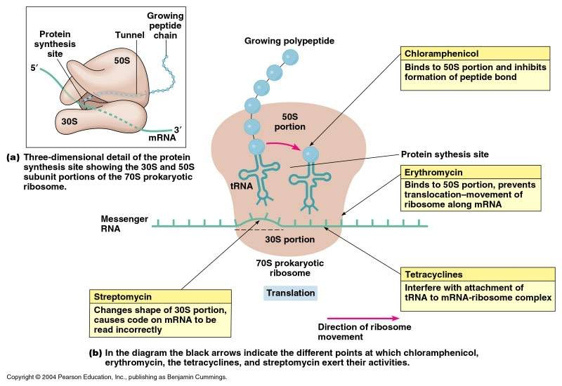
Ribosomes are the workhorses of protein biosynthesis, the process of translating mRNA into protein. The mRNA comprises a series of codons that dictate to the ribosome the sequence of the amino acids needed to make the protein. Using the mRNA as a template, the ribosome traverses each codon (3 nucleotides) of the mRNA, pairing it with the appropriate amino acid provided by a tRNA. Molecules of transfer RNA (tRNA) contain a complementary anticodon on one end and the appropriate amino acid on the other. The small ribosomal subunit, typically bound to a tRNA containing the amino acid methionine, binds to an AUG codon on the mRNA and recruits the large ribosomal subunit. The ribosome then contains three RNA binding sites, designated A, P, and E. The A site binds an aminoacyl-tRNA (a tRNA bound to an amino acid); the P site binds a peptidyl-tRNA (a tRNA bound to the peptide being synthesized); and the E site binds a free tRNA before it exits the ribosome. Protein synthesis begins at a start codon AUG near the 5' end of the mRNA. mRNA binds to the P site of the ribosome first. The ribosome is able to identify the start codon by use of the Shine-Dalgarno sequence of the mRNA in prokaryotes and Kozak box in eukaryotes.

|