CSS Forums
Saturday, April 20, 2024
04:59 AM (GMT +5)
04:59 AM (GMT +5)


|
#101
 Friday, May 29, 2009
|
|||
|
|||
|
Importance of birds Birds are excellent natural indicators of the health of many ecosystems. They are the literal canary in the coalmine--when birds disappear from an area, it normally signals the deteriorating health of the entire ecosystem. The Importance of Birds Birds eat insects. They are a natural way to control pests in gardens, on farms, and other places. They aid in the pollinization of plants. By landing on a plant or sucking the nectar from a flower, and then moving on to the next, a bird does the job usually associated with bees. Birds also have a good system for spreading seeds. They eat berries and then when they "dispose of" their waste, the berry seeds are disposed along with it. Bird feces provide good fertilization for the seeds with which they are dropped, giving seeds very good conditions with which to grow. Above all things, what birds gave us was the dream of flying. Perhaps without birds, we never would have imagined the possibility of flight. Ecological Importance Spring would not be spring without bird songs. Theodore Roosevelt Wild birds are essential components of healthy, functioning natural systems. They pollinate flowers, disseminate seeds, and help keep insect populations under control. Like the proverbial canaries in a coalmine, birds also serve as indicators of the ecological health of our planet. Because of their rapid metabolism and wide geographic distribution, birds provide early warning to us of changes in the environment and potentially harmful biological conditions. Robust, diverse bird populations reflect the underlying health of the ecosystem in which theyand welive. When a wetland begins to lose its ducks, herons, and swallows, its a signal that water quantity and quality are declining, which bodes ill for other species such as salmon and people. Since we share our planet with all other species, what happens to birds will happen to us. Economic Importance Kill not the goose that lays the golden egg. English proverb Birds save money. Without the environmental assistance we get from birds, we would have to spend far more money on pest control and keeping natural systems in balance. Insect-eating birds on farms, in woodlands, and in cities reduce the need for chemical pest control. Birds are also voracious eaters of weed plants and nuisance rodents. They provide us with free ecological services and are unheralded assistants to farmers, foresters, and gardeners. Birds make money. Bird watching is the fastestgrowing form of outdoor recreation in the United States, up 155 percent in the last ten years. In 2001 more than 46 million Americans watched birds and spent more than $32 billion on the pastime. People travel to see birds, buy backyard bird feeders, plant gardens for birds, and spend money to support bird research and protect bird habitat. Birders attract money to Washington. The economic impact from birders and watchable wildlife enthusiasts is astounding. The latest report from the Washington Department of Fish and Wildlife shows that wildlife watching provides more than 22,000 jobs and brings in $980 million to the state annually, compared to $350 million from hunting and $854 million from recreational fishing. Most birders are well educated, have annual incomes greater than $30,000, and are willing to spend money to watch birds. When birders go on overnight bird-watching trips, they typically spend $100 to $130 per day. Aesthetic Importance Keep a green bough in your heart and the singing bird will come. Chinese proverb Birds populate our fields and forests and waters as well as our books and plays and paintings. They awaken the poet and artist and philosopher in each of us, and we celebrate their beauty and power of flight. They help our spirits soar, our visions broaden and they renew our natural sense of joy and wonder. Birds of prey are sometimes accused of killing farm animals, such as chickens. The numbers of farm animals killed by birds of prey is of minor economic consequence when compared to their contributions to pest control.Peregrine falcons (and predatory birds in general) are a great asset to many farmers, killing millions of crop-destroying vertebrates and insects. Valuable Birds Birds are most useful to humans as destroyers of harmful insects and as consumers of weed seeds. Predatory birds such as the hawk, eagle, and owl are essential because they keep down the populations of rats, mice, and other rodents that would otherwise devour valuable food crops. Birds also pollinate many species of flowering plants. Guano is a valuable fertilizer obtained from the droppings of seabirds. Seeds that pass undigested through the bodies of birds fall to the ground and introduce plants into new areas. Domestic birds such as the chicken, duck, turkey, and goose contribute meat and eggs to our food supply. Game birds furnish both meat and sport. The feathers of the ostrich, pheasant, goose, and other species are used for decoration. Eiderdown, duck, goose, and chicken feathers are used to stuff pillows, quilts, and outdoor clothing. Birds such as the canary, parakeet, parrot, and cockatoo are tamed for their singing or "talking" ability. Pigeons are used for racing and as message carriers, and are kept as pets and for food purposes. Various species of falcons and hawks are trained to catch birds and small animals for their masters. In China, cormorants are used to catch fish. Bird Pests In cities, the English sparrow, pigeon, and starling are often annoying because of their droppings and noise. Crows, blackbirds, starlings, and others sometimes destroy plant seedlings. Many kinds of birds attack ripe fruit; and finches, linnets, and pine siskins have been known to destroy crops of cherries, apricots, and peaches by eating the fruit buds. Birds are carriers of the disease psittacosis (parrot fever), which can affect humans. Quarantine and inspection of imported birds have all but eliminated the disease from the United States 
|


| The Following 2 Users Say Thank You to AFRMS For This Useful Post: | ||
abrarchatha (Monday, September 16, 2013), goswamiraaj (Saturday, July 02, 2011) | ||
|
#102
Friday, May 29, 2009
|
||||
|
||||
|
Asalam o Alaikum AFRMS
u r effort is great. i want to get some material on vertebral column of humans , its anatomy and fuction and types of nerves arising from it. if u have some matter plz share it with me if u have some time. i will be thankful to you.
|
|
#103
Friday, May 29, 2009
|
|||
|
|||
|
@ Andy Pandy
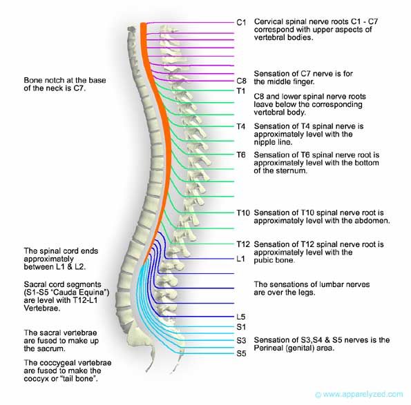
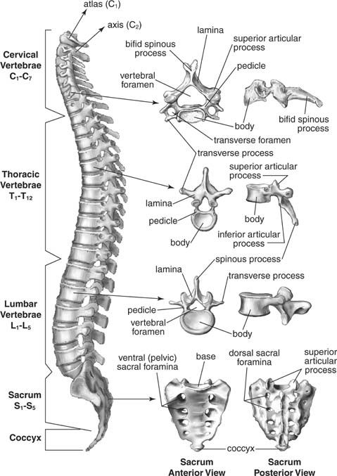
Human Vertebral System In vertebrate animals, the flexible column extending from neck to tail, made of a series of bones, the vertebrae. The major function of the vertebral column is protection of the spinal cord; it also provides stiffening for the body and attachment for the pectoral and pelvic girdles and many muscles. In humans an additional function is to transmit body weight in walking and standing. Each vertebra, in higher vertebrates, consists of a ventral body, or centrum, surmounted by a Y-shaped neural arch. The arch extends a spinous process (projection) downward and backward that may be felt as a series of bumps down the back, and two transverse processes, one to either side, which provide attachment for muscles and ligaments. Together the centrum and neural arch surround an opening, the vertebral foramen, through which the spinal cord passes. The centrums are separated by cartilaginous intervertebral disks, which help cushion shock in locomotion. Vertebrae in lower vertebrates are more complex, and the relationships of their parts to those of higher animals are often unclear. In primitive chordates (e.g., amphioxus, lampreys) a rodlike structure, the notochord, stiffens the body and helps protect the overlying spinal cord. The notochord appears in the embryos of all vertebrates in the space later occupied by the vertebral bodiesin some fish it remains throughout life, surrounded by spool-shaped centrums; in other vertebrates it is lost in the developed animal. In primitive chordates the spinal cord is protected dorsally by segmented cartilagesthese foreshadow the development of the neural arch of true vertebrae. Fish have trunk and caudal (tail) vertebrae; in land vertebrates with legs, the vertebral column becomes further subdivided into regions in which the vertebrae have different shapes and functions. Crocodilians and lizards, birds, and mammals demonstrate five regions: (1) cervical, in the neck, (2) thoracic, in the chest, which articulates with the ribs, (3) lumbar, in the lower back, more robust than the other vertebrae, (4) sacral, often fused to form a sacrum, which articulates with the pelvic girdle, (5) caudal, in the tail. The atlas and axis vertebrae, the top two cervicals, form a freely movable joint with the skull. The numbers of vertebrae in each region and in total vary with the species. Snakes have the greatest number, all very similar in type. In turtles some vertebrae may be fused to the shell (carapace); in birds all but the cervical vertebrae are usually fused into a rigid structure, which lends support in flight. Most mammals have seven cervical vertebrae; size rather than number account for the variations in neck length in different species. Whales show several specializationsthe cervical vertebrae may be either much reduced or much increased in number, and the sacrum is missing. Humans have 7 cervical, 12 thoracic, 5 lumbar, 5 fused sacral, and 3 to 5 fused caudal vertebrae (together called the coccyx). The vertebral column is characterized by a variable number of curves. In quadrupeds the column is curved in a single arc (the highest portion occurring at the middle of the back), which functions somewhat like a bow spring in locomotion. In humans this primary curve is modified by three more: (1) a sacral curve, in which the sacrum curves backward and helps support the abdominal organs, (2) an anterior cervical curve, which develops soon after birth as the head is raised, and (3) a lumbar curve, also anterior, which develops as the child sits and walks. The lumbar curve is a permanent characteristic only of humans and their bipedal forebears, though a temporary lumbar curve appears in other primates in the sitting position. The cervical curve disappears in humans when the head is bent forward but appears in other animals as the head is raised. Human skeletal system The vertebral column is not actually a column but rather a sort of spiral spring in the form of the letter S. The newborn child has a relatively straight backbone. The development of the curvatures occurs as the supporting functions of the vertebral column in humansi.e., holding up the trunk, keeping the head erect, serving as an anchor for the extremitiesare developed. The S-curvature enables the vertebral column to absorb the shocks of walking on hard surfaces; a straight column would conduct the jarring shocks directly from the pelvic girdle to the head. The curvature meets the problem of the weight of the viscera. In an erect animal with a straight column, the column would be pulled forward by the viscera. Additional space for the viscera is provided by the concavities of the thoracic and pelvic regions. Weight distribution of the entire body is also effected by the S-curvature. The upper sector to a large extent carries the head; the central sector carries the thoracic viscera, the organs and structures in the chest; and the lower sector carries the abdominal viscera. If the column were straight, the weight load would increase from the head downward and be relatively great at the base. Lastly, the S-curvature protects the vertebral column from breakage. The doubly bent spring arrangement is far less vulnerable to fracture than would be a straight column. The protective function of the skeleton is perhaps most conspicuous in relation to the central nervous system, although it is equally important for the heart and lungs and some other organs. A high degree of protection for the nervous system is made possible by the relatively small amount of motion and expansion needed by the component parts of this system and by certain physiological adaptations relating to circulation, to the cerebrospinal fluid, and to the meninges, the coverings of the brain and spinal cord. The brain itself is snugly enclosed within the boxlike cranium. Sharing in the protection afforded by the cranium is the pituitary gland, or hypophysis. In human anatomy, the vertebral column (backbone or spine) is a column usually consisting of 24 vertebrae,[1] the sacrum, intervertebral discs, and the coccyx situated in the dorsal aspect of the torso, separated by spinal discs. It houses the spinal cord in its spinal canal. Curves Viewed laterally the vertebral column presents several curves, which correspond to the different regions of the column, and are called cervical, thoracic, lumbar, and pelvic. The cervical curve, convex forward, begins at the apex of the odontoid (tooth-like) process, and ends at the middle of the second thoracic vertebra; it is the least marked of all the curves. The thoracic curve, concave forward, begins at the middle of the second and ends at the middle of the twelfth thoracic vertebra. Its most prominent point behind corresponds to the spinous process of the seventh thoracic vertebra. This curve is known as a tt curve. The lumbar curve is more marked in the female than in the male; it begins at the middle of the last thoracic vertebra, and ends at the sacrovertebral angle. It is convex anteriorly, the convexity of the lower three vertebrae being much greater than that of the upper two. This curve is described as a lordotic curve. The pelvic curve begins at the sacrovertebral articulation, and ends at the point of the coccyx; its concavity is directed downward and forward. The thoracic and pelvic curves are termed primary curves, because they alone are present during fetal life. The cervical and lumbar curves are compensatory or secondary, and are developed after birth, the former when the child is able to hold up its head (at three or four months) and to sit upright (at nine months), the latter at twelve or eighteen months, when the child begins to walk. Names of individual vertebrae Individual vertebrae named according to region and position, from superior to inferior
|
|
#104
Saturday, May 30, 2009
|
|||
|
|||
|
Bird Migration Migration occurs wherever there are birds. Migration is the part of the animals life that is characterized by geographic movements. This is an incredibly diverse behavior, an adaptation that has been shaped by natural selection. Arctic birds migrate, as do tropical birds. We often think of migration as the seasonal movement of birds during spring and fall to avoid harsh winters. We have all heard the phrase fly south for the winter. This is only partly correct. Scientists have extensively studied the evolution of migration. Not one theory has been widely accepted. Explanations include and are not limited to glaciers, changing climatic patterns, and extending dispersal distance. Most biologists will agree that the foundations of all explanations must be food. The availability of food is the driving force in the evolution of migration. Food abundance can increase reproductive output, whereas a lack of food leads to death a bird that can find food will have a longer life span and produce offspring more readily than one that finds less food. Just by moving from place to place a bird can find more food, and thus migration evolved. It just so happens that many warmer climates have an overabundance of food, whereas cold harsh environments do not. Most species of birds can tolerate colder temperatures given that food is available. Scientists have identified three patterns of migration: complete, partial and irruptive. Complete migration occurs when individuals leave the breeding range during the no breeding season. Many North American birds such as warblers, orioles, hummingbirds, and shorebirds exhibit this migration pattern. Complete migrants may travel incredible distances, sometimes more than 15,000 miles per year. The most common type of migration is partial migration. This is characterized by seasonal movements away from a breeding range by some but not all of the members of a species. For example, Song Sparrows migrate south for the winter, but some individuals remain in the breeding area. Thus the migration is not complete across the species, instead it is only partial. Partial migrants, like complete migrants, take advantage of seasonally abundant food. Migrations that are not seasonally or geographically predictable are said to be irruptive. Such migrations may occur one year but not again for many years. The distances and numbers of individuals involved are also less predictable than with complete or partial migrants. The Great Gray Owl is an irruptive migrant. It migrates south only occasionally and the number of these owls that migrate vary. Many scientists call these irruptive migrants food specialists. Many of these migrants may only eat certain seeds from certain trees. When the seeds are available, irruptive migrants stay; when the seeds are difficult to find, they move. The factors that are believed to control the onset of migration can be divided roughly into external (exogenous) or internal (endogenous) factors. Both at different times can control the seasonal timing of migration. Migration onset and seasonal timing are controlled genetically in some species. This would be an example of an endogenous factor. In other species, the onset of migration is controlled by weather, food, or social factors. These would be examples of exogenous factors. It is difficult to study endogenous factors such as genetic control. How do birds find there way to their destination? Studies have shown that migratory birds find their way over long distances, which implies that these birds must have a sort of mental compass and map. The migratory pathways used by birds are influenced by topography, geography and wind. These routes are not always the same from year to year. Bodies of water, deserts, and mountain ranges are the physiographic features of the earth that can act as barriers. Many researchers have concluded that wind is one of the strongest factors in determining a migration pattern. Many birds rely on the direction of the wind to get them there. This is also true in airplanes. If the wind is in your favor, your flight will be faster. Research has revealed that many migratory birds take advantage of winds to make better progress. Many scientists believe that a bird has an internal compass that permits orientation in a given direction. Many researchers have thought that birds rely on olfactory or magnetic cues to guide them to their destinations. Another hypothesis suggests that birds have an innate time program coupled to an innate flight direction. Birds are thought to be genetically programmed to fly in a given direction for a given period of time. Some studies have proven this theory to be correct; only more research and time will tell. How do scientists study migration? One of the most popular methods of studying migration is banding birds. Banding is a four-part process. First scientists catch the birds. Then they band the birds (usually a leg band encoded with a number and other information). They then release the birds into the wild. Finally they transfer the information into a database. Banding does have its shortcomings. For starters, most birds that are banded are never seen again. Birds are hard to track. Another method that has been used is radar. Most radar is used to examine the movement of large numbers of birds overall several hundred square kilometers. The position of each bird can be recorded every few seconds and its speed, altitude, direction and mode of light are measured with precision. Radar is a wonderful tool and the only shortcoming appears to be that it is effective for only a small area. Radiotelemetry is another technique. This involves putting radiotransmitters on birds so that their movements and activities can be monitored from a distance. The transmitters weigh less than three percent of a birds body weight, and birds tend to adjust to them rapidly. The transmitter can send out a signal every few seconds. Most transmitters are battery powered, but some are solar powered. Once a transmitter is attached to a bird, the researcher follows it from an automobile or airplane fitted with an antenna to receive the transmitted signal. Data gathered through radiotelemetry is more detailed than other methods of study. The physiological and neural mechanisms that control a migrants behavior can be studied in migration laboratories that are designed to duplicate the migratory environment. A researcher will often create a controlled environment that stimulates one or more aspects of migration, than manipulates them experimentally. Orientation, navigation, and biological rhythms are the most often studied in laboratories. All such methods are extremely helpful in uncovering the mystery of migration. Even though much research has been done in this field, much more still needs to be completed. There are many unknowns when it comes to migration. Most of the information we have is based on theories that have been demonstrated experimentally. This makes it hard to imagine any such theory on migration being set in stone. Migratory behaviors and routes can change and this is why it is often frustrating for scientists. The next time you see a bird flying overhead, you may ponder where they are going and how many miles they will travel in order to get there. Navigation How do birds find their way? Simple. Through a combination of · Sighting (they don't call it a "bird's eye view" for nothing) features like rivers, coastlines, and mountain ranges. · Monitoring Earth's magnetic field, apparently with their visual system and with tiny grains of a mineral called magnetite in their heads · Observing the stars · Using the sun for guidance · Smell · And probably following their neighbors (many birds migrate in large flocks) Question: does it still sound simple? I didn't think so. For example, birds, which use magnetic navigation, must deal with a small problem -- magnetic north is 1,600 kilometers from the North Pole. That means migrants leaving northern Alaska and following magnetic south would be traveling due west! Second, star navigation changes as new constellations appear on the horizon as the birds travel north or south. Although birds have apparently been overcoming these problems for millions of years, only in the past year have scientists figured out how at least one species of bird does it (see Constant Compass Calibration). It seems that the birds recalibrate their magnetic compasses against their star navigation during their rest stops along the migration route. And if they don't have enough time at the rests, they get lost. Flight strategies Once the birds know where they're going, they also need a way to get there, and that brings us to the realm of fuel efficiency -- of miles per rodent, or kilometers per thistle seed. Depending on their size, route, and laziness (just kidding!), birds use one of these flight strategies: · Slow and Steady Wins the Race: The most basic technique is to keep flapping your wings until you land. That's the technique used by the Canada goose and many other migrants. · Soaring. Smarter (still kidding) birds have figured out how to ride "thermals" -- updrafts of air caused by solar heating, to take a free ride high into the sky. These birds, including Swainson's hawk, turkey vultures, and many others, only travel during the day, and only over land (preferably flat land). Those restrictions can lead to astonishing concentrations of migrating birds. · Flapping and gliding. These birds flap their wings for a few beats, and then glide for a while. After they lose some altitude and/or speed, they flap some more. · Bounding. This is a combination of flapping with a closed-wing glide. Birds whose wings would produce too much drag use it. Although their aerodynamic bodies do create some lift, the birds tend to lose altitude and the flight pattern is up-and-down, like the flap-and-glide pattern. Rapid Transit: the ins and outs of bird migration Let's face it--when it comes to dealing with winter, most birds seem an awful lot smarter than humans. Instead of griping about the weather, they simply head for a warmer climate. Let's look at a few facts on bird migration: · What's the record for the longest migration on the planet? The arctic tern flies a phenomenal round trip that can be as long as 20,000 miles per year, from the Arctic to the Antarctic and back. Other sea birds also make astounding journeys: the long-tailed Jaeger flies 5,000 to 9,000 miles in each direction. · The sand hill and whooping cranes are both capable of migrating as far as 2.500 miles per year, and the barn swallow more than 6,000 miles. · Why do about 520 of the 650 bird species that nest in the United States migrate south to spend the winter? Because they get bored shivering in the dark. And because it's bleak in the winter. And because there's nothing to eat. And because their ancestors did it. · Why do some birds go north for the summer? Because there's more to eat. The 24-hour days near the Arctic Circle produces a fantastic flowering of life. This brief, but abundant, source of food attracts many birds (and mammals such as the caribou) to the Arctic for breeding purposes. · What influences migration patterns over the long term? Changes in climate (particularly ice ages), and shifts in the positions of islands and continents as a result of tectonic drift. · How do they keep going? Some birds store a special, high-energy fat before the trip. Soaring raptors, for example, may not eat for several weeks as they migrate. Other species eat along their migration routes. · How high can they fly? Higher than Mt. Everest. Bar-headed geese have been recorded flying across the Himalayas at 29,000 feet. Other species seen above 20,000 feet include the whooper swan, the bar-tailed godwit, and the mallard duck. (Note: birds don't fly this high just to get in the Guinness book of records, but rather to reach their destinations efficiently. From radar studies, scientists know that birds can change altitudes to find the best wind conditions. To fight a headwind, most birds stay low, where ridges, trees and buildings slow the wind. To ride a tailwind, they get up high where the wind is as fast as possible.) The world's largest migration of raptors (hawks, vultures, falcons, kites and other species) flies across Mexico's Caribbean coastal plain near the city of Vera Cruz. Raptor watching is an old specialty among birders--and thousands of eager birders flock to Cape May, New Jersey to watch 50,000 of these elegant fliers make their way between wintering and breeding grounds. In biology, the term migration refers to the regular, periodic movement of animals between two different places. Migration usually occurs in response to seasonal changes and is motivated by breeding and/or feeding drives. Migration has been studied most intensively among birds, but it is known to take place in many other animals as well, including insects, fish, whales, and other mammals. Migration is a complex behavior that involves timing, navigation, and other survival skills. The term migration also applies to the movement of humans from one country to another for the purpose of taking up long-term or permanent residency in the new country. Types of migration Four major types of migration are known. In complete migration, all members of a population travel from their breeding habitat at the end of that season, often to a wintering site hundreds or even thousands of kilometers away. The arctic tern is an example of a complete migrant. Individuals of this species travel from the Arctic to the Antarctic and back again during the course of a year, a round-trip migration of more than 30,000 kilometers! In other species, some individuals remain at the breeding ground year-round while other members of the same species migrate away. This phenomenon is known as partial migration. American robins are considered indicators of the arrival of spring in some areas but are year-round residents in other areas. Differential migration occurs when all the members of a population migrate, but not necessarily at the same time or for the same distance. The differences are often based on age or sex. Herring gulls, for example, migrate a shorter and shorter distance as they grow older. Male American kestrels spend more time at their breeding grounds than do females, and when they do migrate, they don't travel as far. Irruptive migration occurs in species that do not migrate at all during some years but may do so during other years. The primary factors determining whether or not migration occurs are weather and availability of food. For example, some populations of blue jays are believed to migrate only when their winter food of acorns is scarce. Migration Pathways Migratory animals travel along the same general routes each year. Several common "flyways" are used by North American birds on their southward journey. The most commonly used path includes an 800 to 1,100 kilometer flight southward across the Gulf of Mexico. In order to survive this difficult journey, birds must store extra energy in the form of fat. All along the migration route, but particularly before crossing a large expanse of water, birds rest and eat, sometimes for days at a time. The birds start out again on their journey only when they have added a certain amount of body fat. Although most migrants travel at night, a few birds prefer daytime migrations. The pathways used by these birds tend to be less direct and slower than those of night migrants, primarily because of differences in feeding strategies. Night migrants can spend the day in one area foraging for food and building up energy reserves for the night's nonstop flight. Daytime migrants must combine travel with foraging, and thus tend to keep to the shorelines, which are rich in insect life, capturing food during a slow but ever-southward journey. Navigation Perhaps the most remarkable aspect of migration is the navigational skills employed by the animals. Birds such as the albatross and lesser golden plover travel hundreds of kilometers over the featureless open ocean. Yet they arrive home without error to the same breeding grounds year after year. Salmon migrate upstream from the sea to the very same freshwater shallows in which they were hatched. Monarch butterflies began life in the United States or Canada. They then travel to the same wintering grounds in Southern California or Mexico that had been used by ancestors many generations before. How are these incredible feats of navigation accomplished? Different animals have been shown to use a diverse range of navigational aids, involving senses often much more acute than our own. Sight, for example, may be important for some animals' navigational skills, although it may often be secondary to other senses. Salmon can smell the water of their home rivers, and follow this scent all the way from the sea. Pigeons also sense wind-borne odors and may be able to organize the memories of the sources of these smells in a kind of internal map. It has been shown that many animals have the ability to sense the magnetic forces associated with the north and south poles, and thus have their own built-in compass. This magnetic sense and the sense of smell are believed to be the most important factors involved in animal migration. Migration Adaptations Many types of birds migrate -- songbirds, raptors, shorebirds and waterfowl. There are as many styles of migration as there are types of birds. Some birds migrate during the day while others fly at night. Some fly alone while others flock together. Some flocks are highly ordered while others are loose congregations. Some birds travel at high altitudes while others fly low. Some fly over water while others stay over land. Migration is one of the most widely studied areas of bird biology, yet it is still poorly understood. People have observed and noted migrations for centuries, yet we still have only a handful of theories about how birds accomplish such impressive flights each year. Migration seems to be triggered by a combination of shortening days and seasonal atmospheric changes, which allow cold fronts to carry frigid air on strong winds. As temperatures drop, insects and plants die while amphibians and rodents hibernate, causing food sources to become scarce. To survive the cold and the competition, birds alight on the strong southerly blowing winds. As autumn progresses, millions of birds disperse to the reaches of the globe to find the food they need to survive. In addition to triggering atmospheric changes, shortening days cause physiological and behavioral changes in the birds. To prepare for migration, birds literally gorge themselves. Fat fuels their flight. 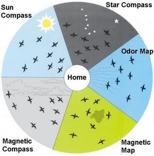
|
|
#105
Saturday, May 30, 2009
|
|||
|
|||
|
Respiration in Mammals To provide the gas exchange necessary to support the elevated metabolic rate of mammals, mammalian lungs are subdivided internally. The repetitive subdivisions of the lung airways provide gas to the tiny alveoli (gas sacs) that form the functional gas-exchange surface area of the lungs. Human lungs have an estimated 300,000,000 alveoli, providing in an adult a total surface area approximately equivalent to a tennis court. Inspiration in mammals, as in reptiles, is powered by an aspiration (suction) pump. Expansion of the chest lowers the pressure between the lungs and the chest wall, as well as the pressure within the lungs. This causes atmospheric air to flow into the lungs. The chief muscles of inspiration are the diaphragm and the external intercostal muscles. The diaphragm is a domelike sheet of muscle separating the abdominal and chest cavities that moves downward as it contracts. The downward motion enlarges the chest cavity and depresses the organs below. As the external intercostal muscles contract, the ribs rotate upward and laterally, increasing the chest circumference. During severe exercise other muscles may also be used. Inspiration ends with the closing of the glottis. In expiration, the glottis opens, and the inspiratory muscles relax; the stored energy of the chest wall and lungs generates the motive power for expiration. During exercise or when respiration is laboured, the internal intercostal muscles and the abdominal muscles are activated. The internal intercostals produce a depression of the rib cage and a decrease in chest circumference. Closely coupled with the circulatory system is the ventilatory (breathing) apparatus, the lungs and associated structures. Ventilation in mammals is unique. The lungs themselves are less efficient than those of birds, for air movement consists of an ebb and flow, rather than a one-way circuit, so a residual volume of air always remains that cannot be expired. Ventilation in mammals is by means of a negative pressure pump made possible by the evolution of a definitive thoracic cavity with a diaphragm. The diaphragm is a unique composite structure consisting of (1) the transverse septum (a wall that primitively separates the heart from the general viscera); (2) pleuroperitoneal folds from the body wall; (3) mesenteric folds; and (4) axial muscles inserting on a central tendon, or diaphragmatic aponeurosis. The lungs lie in separate airtight compartments called pleural cavities, separated by the mediastinum. As the size of the pleural cavity is increased, the lung is expanded and air flows in passively. Enlargement of the pleural cavity is produced by contraction of the diaphragm or by elevation of the ribs. The relaxed diaphragm domes upward, but when contracted it stretches flat. Expiration is an active movement brought about by contraction of abdominal muscles against the viscera. Air typically enters the respiratory passages through the nostrils, where it may be warmed and moistened. It passes above the bony palate and the soft palate and enters the pharynx. In the pharynx the passages for air and food cross. Air enters the trachea, which divides at the level of the lungs into primary bronchi. A characteristic feature of the trachea of many mammals is the larynx. Vocal cords stretch across the larynx and are vibrated by forced expiration to produce sound. The laryngeal apparatus may be greatly modified for the production of complex vocalizations. In some groupsfor example, howler monkeysthe hyoid apparatus is incorporated into the sound-producing organ, as a resonating chamber. Physiology of respiratory system in mammals For more detailed descriptions see also Respiratory physiology or Respiration. Ventilation Ventilation of the lungs is carried out by the muscles of respiration. Control Ventilation occurs under the control of the autonomic nervous system from parts of the brain stem, the medulla oblongata and the pons. This area of the brain forms the respiration regulatory center, a series of interconnected brain cells within the lower and middle brain stem which coordinate respiratory movements. The sections are the pneumotaxic center, the apneustic center, and the dorsal and ventral respiratory groups. This section is especially sensitive during infancy, and the neurons can be destroyed if the infant is dropped and/or shaken violently. The result can be death due to "shaken baby syndrome." Inhalation Inhalation is initiated by the diaphragm and supported by the external intercostal muscles. Normal resting respirations are 10 to 18 breaths per minute, with a time period of 2 seconds. During vigorous inhalation (at rates exceeding 35 breaths per minute), or in approaching respiratory failure, accessory muscles of respiration are recruited for support. These consist of sternocleidomastoid, platysma, and the scalene muscles of the neck. Under normal conditions, the diaphragm is the primary driver of inhalation. When the diaphragm contracts, the ribcage expands and the contents of the abdomen are moved downward. This results in a larger thoracic volume and negative (suction) pressure (with respect to atmospheric pressure) inside the thorax. As the pressure in the chest falls, air moves into the conducting zone. Here, the air is filtered, warmed, and humidified as it flows to the lungs. During forced inhalation, as when taking a deep breath, the external intercostal muscles and accessory muscles aid in further expanding the thoracic cavity. Exhalation Exhalation is generally a passive process; however, active or forced exhalation is achieved by the abdominal and the internal intercostal muscles. During this process air is forced or exhaled out. The lungs have a natural elasticity: as they recoil from the stretch of inhalation, air flows back out until the pressures in the chest and the atmosphere reach equilibrium.[7] During forced exhalation, as when blowing out a candle, expiratory muscles including the abdominal muscles and internal intercostal muscles, generate abdominal and thoracic pressure, which forces air out of the lungs. Circulation The right side of the heart pumps blood from the right ventricle through the pulmonary semilunar valve into the pulmonary trunk. The trunk branches into right and left pulmonary arteries to the pulmonary blood vessels. The vessels generally accompany the airways and also undergo numerous branchings. Once the gas exchange process is complete in the pulmonary capillaries, blood is returned to the left side of the heart through four pulmonary veins, two from each side. The pulmonary circulation has a very low resistance, due to the short distance within the lungs, compared to the systemic circulation, and for this reason, all the pressures within the pulmonary blood vessels are normally low as compared to the pressure of the systemic circulation loop. Gas exchange The major function of the respiratory system is gas exchange between the external environment and an organism's circulatory system. In humans and mammals, this exchange facilitates oxygenation of the blood with a concomitant removal of carbon dioxide and other gaseous metabolic wastes from the circulation. As gas exchange occurs, the acid-base balance of the body is maintained as part of homeostasis. If proper ventilation is not maintained, two opposing conditions could occur: 1) respiratory acidosis, a life threatening condition, and 2) respiratory alkalosis. Upon inhalation, gas exchange occurs at the alveoli, the tiny sacs which are the basic functional component of the lungs. The alveolar walls are extremely thin (approx. 0.2 micrometres). These walls are composed of a single layer of epithelial cells (type I and type II epithelial cells) in close proximity to the pulmonary capillaries which are composed of a single layer of endothelial cells. The close proximity of these two cell types allows permeability to gases and, hence, gas exchange. Non-respiratory functions Vocalization The movement of gas through the larynx, pharynx and mouth allows humans to speak, or phonate. Vocalization, or singing, in birds occurs via the syrinx, an organ located at the base of the trachea. The vibration of air flowing across the larynx (vocal chords), in humans, and the syrinx, in birds, results in sound. Because of this, gas movement is extremely vital for communication purposes. Temperature control Panting in dogs and some other animals provides a means of controlling body temperature. This physiological response is used as a cooling mechanism. Coughing and sneezing Irritation of nerves within the nasal passages or airways, can induce coughing and sneezing. These responses cause air to be expelled forcefully from the trachea or nose, respectively. In this manner, irritants caught in the mucus which lines the respiratory tract are expelled or moved to the mouth where they can be swallowed. Development of respiratory system in animals Humans and mammals The respiratory system lies dormant in the human fetus during pregnancy. At birth, the respiratory system becomes fully functional upon exposure to air, although some lung development and growth continues throughout childhood. Pre-term birth can lead to infants with under-developed lungs. These lungs show incomplete development of the alveolar type II cells, cells that produce surfactant. The lungs of pre-term infants may not function well because the lack of surfactant leads to increased surface tension within the alveoli. Thus, many alveoli collapse such that no gas exchange can occur within some or most regions of an infant's lungs, a condition termed respiratory distress syndrome. Basic scientific experiments, carried out using cells from chicken lungs, support the potential for using steroids as a means of furthering development of type II alveolar cells.[8]In fact, once a pre-mature birth is threatened, every effort is made to delay the birth, and a series of steroid shots is frequently administered to the mother during this delay in an effort to promote lung growth Mammals For mammals, including humans, respiration is essential. In these organisms, the respiratory system can be subdivided into an upper respiratory tract and a lower respiratory tract based on anatomical features. The upper respiratory tract includes the nasal passages, pharynx and the larynx, while the lower respiratory tract is comprised of the trachea, the primary bronchi and lungs. The respiratory system can also be divided into physiological, or functional, zones. These include the conducting zone (the region for gas transport from the outside atmosphere to just above the alveoli), the transitional zone, and the respiratory zone (the alveolar region where gas exchange occurs). (See also respiratory tract.) Comparative anatomy/physiology in mammals Horses Horses are obligate nasal breathers. That is, they are different from many other mammals in that they do not have the option of breathing through their mouths and must take in air through their nose. Elephants The elephant is the only mammal known to have no pleural space. Rather, the parietal and visceral pleura are both composed of dense connective tissue and joined to each other via loose connective tissue. This lack of a pleural space, along with an unusually thick diaphragm, are thought to be evolutionary adaptations allowing the elephant to remain underwater for long periods of time while breathing through its trunk which emerges as a snorkel. 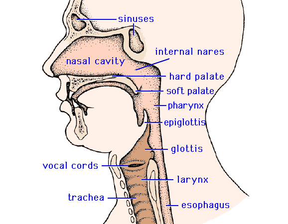 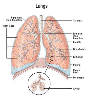  Last edited by Viceroy; Saturday, May 30, 2009 at 08:35 PM. Reason: Merger
|
|
#106
Wednesday, June 03, 2009
|
|||
|
|||
|
Mammal Dentition Teeth are important things, without them eating becomes a lot more difficult. In mammals teeth have reached their highest peak of evolution, mammalian teeth are both more complicated and more efficient than in other vertebrates. Teeth are heavy and require considerable muscle to operate efficiently, this has made an important contribution to the evolution of the mammal skull. Mammals are heterodonts, this means some of our teeth are different. In fishes and reptiles the teeth are all basically the same, some bigger than others but the same basic shape. Mammals needed their teeth to do several different jobs and so mammal teeth evolved into different forms. Mammal teeth can grind, stab, scissor, dig, chisel, sieve and lift (elephants tusks). Teeth are the hardest part of any mammal and therefore they are the part most often fossilised. The number, size, organisation and shape of the teeth are different in every species of mammal and can be used in taxonomy, especially of fossils. In fact without teeth the fossil record would be much harder to understand. Mammals have only two sets of teeth, the first set they get soon after birth, often called the 'milk teeth' and a larger set they acquire as an adult. The larger set has both more and larger teeth to fill the larger jawbones In all other toothed vertebrates teeth just keep coming, no matter how many you lose there is always another one ready to take its place. In other words fish amphibians, reptiles and birds either have no teeth or numerous sets. Teeth do not last for ever, like everything they wear out. How fast they wear out depends on what the animal eats. Herbivore teeth in particular tend to where out at a specific rate. This is very useful for biologists as it allows them to age an animal by looking at its teeth. Therefore even a skull of a long dead animal can supply useful information by faithfully retaining the information on how old the animal was when it died. Elephants, as you might have expected have the largest teeth in the world. An Elephants tusks are actually modified incisors. They arise from the upper jaw and only 2/3s of them are visible because they are deeply embedded in the elephants skull. The heaviest pair of tusks, and therefore the heaviest pair of teeth of any extant (still living species) animal belonged to an African Elephant Loxodonta africana shot in 1897. One of the pair was slightly larger than the other but together they weighed 211kg or 465lb. A single tusk from an unknown African Elephant weighed in at 117kg or 258lb Paris in 1900. this is the single largest tooth of a modern animal. Prehistoric records also go to members of the elephant family. The longest tusk ever found belonged to the extinct Palaeoloxodom antiquus germanicus, the Straight-tusked Elephant. On average this magnificent animal had tusks 5m or 16.5ft long. The heaviest tusks ever known belonged to the Colombian Mammoth Mammuthus columbi, these weighed 226kg or 498lb. A single tusk from an unknown species that weighs 150kg or 330lb is preserved in a museum in Milan. If the owner of this huge tooth had two approximately the same size their combined weight would have been around 300kg or 660lb and thus far more than the Colombian Mammoth mentioned first. Apart from elephants the tusks of Walruses can also get quite large being up to 1m long and weighing 5.4kg or 12lb. The canines of hippopotamuses can also be quite impressive. Tusks aside it is the African Elephant again who comes in as the winner in the largest teeth stakes. An bull African Elephant's molars can be easily be more than a foot long and weigh 4kg or 10lb. The number of teeth in an animals jaw depend on its life history. Teeth are expensive to grow so no animal wants more than it needs. Most placental mammals are happy with between 20 and 40, while most marsupials have 30 to 50. As a general rule animals that feed on insects have more teeth than either herbivores or the larger carnivores. There are some funny exceptions though. Several groups of mammals have decided to do without teeth altogether. The 10 species of Whales in the order Mysticeti, the 8 species of Pangolins family Manidae, and the 3 species of Anteaters in the family Myrmecophagidae and order Edentata have all given up on teeth completely and have none. An interesting tooth oddity is the Narwhale Monodon monocerus, order Odontoceti. This has two teeth in its upper jaw, one of these, normally the left one, grows out long and forward as a tusk while the other remains rudimentary. Looking the other way to see who has the most teeth we have to visit two of the 3 orders that gave us the mammals with the least teeth. On land the mammal with the most teeth is the Giant Armadillo Priodontes giganteus order Edentata, which can as many 100 teeth in its jaws. In the oceans the real master of teeth comes from the order Odontoceti. The Long-snouted Spinner Dolphin can have as many as 252 teeth in its long thin jaws. These teeth are more like reptile teeth in that they are all the same basic shape, thin sharp little spears. This makes them good for catching and holding the slippery little fishes that dolphins live on. What is a Tooth Teeth, like bones, are living structures. They rest in a specially designed cavity in the bones of the jaw and at least while they are growing they have a supply of blood and nutrients to them through their base. The crown is the outside of the part of the tooth that is above the jaw bone, it is the part that does all the hard work. It is capped with an extra hard substance called enamel. The rest of the tooth is softer, but still harder than bone. Beneath the enamel, making up the bulk of the tooth is the Dentine and within the centre is the pulp cavity. Enamel is 96% mineral while the dentine is 70% mineral. Teeth are seated in the jawbones and held in place by a special cement which is another form of bone. In the bottom of the tooth is a channel leading between the pulp cavity and the jaw bone which allows blood vessels to access the tooth. Most teeth stop growing once they reach a adult size and the cavity in the bottom of the tooth seals up, but some teeth like the incisors of rodents or the molars of sheep keep growing as they are worn down. Teeth can be divided into two sections. The part above the gum called the crown and the part embedded in the gum called the root. 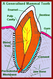 Teeth in mammals come in four different sorts: Incisors, Canines, Premolars and Molars. Not all mammals have all, or even any of them and the roles any particular sort of teeth play in an animal's life can be quite diverse. The arrangement of teeth in any given mammals mouth can be expressed as a 'dental formula'. This formula gives the arrangement of one side of an animal's jaw such that incisors are always written first then canines then premolars and then molars. For humans 2123-2123 is upper and lower jaws respectively signifying 2 incisors, 1 canine, 2 premolars and 3 molars on each side in both the upper and lower jaw. In total this adds up to 8 incisors, 4 canines, 8 premolars and 12 molars = 32 teeth in humans. Other mammals have different numbers of each sort of teeth in their upper and lower jaws. Hence Smoky Bats (family Furipteridae) have the dental formula 2123-3133 whilst Hyenas have 3141-3131 normally. Incisors = Cutting teeth - these are the front most teeth in the jaw primarily used for the initial biting of food. They have a straight, sharp cutting edge and one root. In many Rodents they grow continually throughout the animals life, this is because they get warn down gnawing through things. Canines= Stabbing teeth - normally only 2 pairs (one each side) per jaw. They have a sharp, pointed edge and are used with the incisors to bite into food and or to kill prey. Like incisors they have one root. The tusks of many animals such as elephants are modified canines. They are missing in rodents and most large herbivores (Perissodactyls and Artiodactyls). The gap where the canines would have been is often enlarged and is called a 'diastema'. Premolars= Next back from the canines. They are generally similar to molars in form and function in both herbivores and omnivores, but in carnivores some of them at least are slimmer and are used to cut flesh. When they (the first lower premolar and last upper premolar) are modified like this they are called the carnassials. Otherwise premolars are teeth we use to crush and grind our food. Their upper surfaces have a broad, lumpy top instead of a sharp biting edge. These small irregular lumps are called cusps. Premolars are called bicuspids in some books, this is because, in most cases, they have two cusps. The prefix bi meaning two. The first upper premolars normally have two roots. The other premolars have one root. Molars = These are larger than premolars and extremely variable depending on the animal's diet. Like premolars they are used for crushing and grinding food, and like premolars their upper surfaces have ridges called cusps on them. Molars normally have three to five cusps and two or three roots. In humans we call the third molars, those closest to the back of the mouth 'Wisdom Teeth'. If the jaw bone is not large enough to accommodate all the teeth in it, as sometimes happens with humans, these wisdom teeth can become painfully wedged between the back of the jaw bone and the 2nd molars. This condition is known as 'impacted wisdom teeth'. The human dental formula is: Dentition
2.1.2.3 2.1.2.3 Of cats it is: Dentition 3.1.3.1 3.1.2.1 The attachment of teeth to the bones of the jaw Skin attachment - Teeth are anchored to the skin by collagen fibers that run into the dentine from the dermis (in sharks). Ligamentous in fangs of rattlesnakes Pleurodont - may be ancestral attachment Each tooth touches the bone with the outer surface of the root, may be joined to the jaw by cement or collagenous fibers. Acrodont - scarcely have any roots teeth abut against the rim of the jawbone by a continuous rim of hard tissue (most Osteichthyes). Thecodont - roots of teeth in sockets (alveoli) in jawbone The replacement of teeth in the mouth Most vertebrates replace their teeth continuously, generation after generation, throughout life. Polyphyodont Most mammals have only 2 generations of teeth. Diphyodont Some acrodont teeth and plates of fused teeth are never replaced. Whether the teeth are the same or different in shape Homodont - Teeth are all about the same shape (most vertebrates, few mammals). Heterodont - Teeth have different form and functions in different parts of the tooth row (mammals, a few fish). Two characteristics of cheek teeth: The surface characteristics of the cheek teeth Bunodont: omnivorous, flat crowns covered with enamel Lophodont: transverse ridged cusps Selenodont: longitudinal ridged cusps Secodont: carnassials carnivorous dentition The height of the cheek teeth Brachydont: low crowned Hypsodont: high crowned Examples: Alligator shows dentition that is thecodont (attachment), polyphyodont (replacement), homodont (variety of teeth). Mammals show dentition that is thecodont, heterodont. Mammalian cheek teeth may be designed for crushing and cutting of food - teeth capable of withstanding pressure - or designed to withstand wear. Modifications for crushing / cutting: 1. Low crowned teeth (short and squat) grow to a specific size and then stop. The entire tooth surface is encased in enamel, the strongest of the tooth substances. Such teeth are brachydont. Carnivores have shearing, cutting molars - brachydont, secodont dentition. Epitomized by cat, cougar with single row of cusps Bear, human dentition is omnivorous - brachydont, bunodont dentition Flat crowned, dome like crushing molars Modifications to resist wear: 2. Tall, high crowned hypsodont dentition White - enamel Brown - dentine Enamel wears less than dentine or cement between crowns, leaving a rough, file-like surface. Self-sharpening file Deer - ridges of teeth run anterior-posterior (front to back) moonshaped, hypsodont, selenodont dentition chew back and forth Beaver - hypsodont, Lophodont chew side to side ridges run side to side Horse- hysodont, selenolophodont ridges run in both directions chew in a circle 3. Rootless teeth do have unconstricted roots, grow continuously Rodents such as beaver Heterodont teeth require thecodont attachment. Snake fang attached by a ligament, formed by two fused teeth to create fang. Wolf eel has heterodont, acrodont teeth. 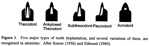 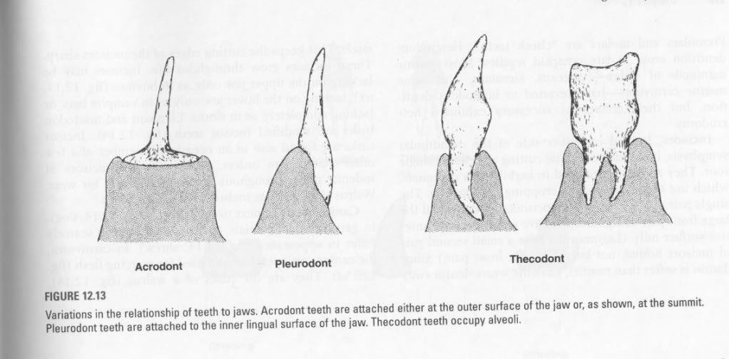 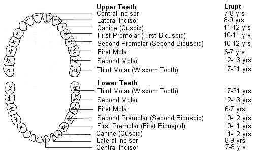 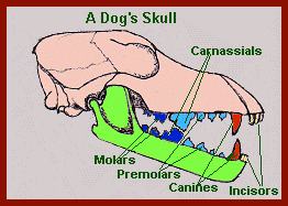
|
|
#108
Friday, June 05, 2009
|
|||
|
|||
|
Plasma Membrane Structure and Function One universal feature of all cells is an outer limiting membrane called the plasma membrane. In addition, all eukaryotic cells contain elaborate systems of internal membranes, which set up various membrane-enclosed compartments within the cell. Cell membranes are built from lipids and proteins. 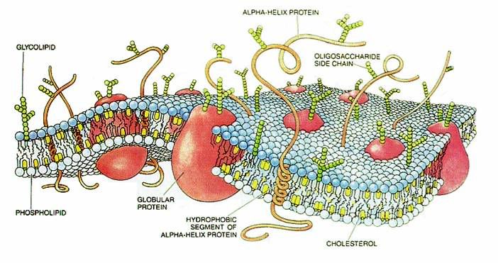 A thin membrane surrounds every living cell, delimiting the cell from the environment around it. The cell membrane surrounds the cytoplasm of a cell and, in animal cells, physically separates the intracellular components from the extracellular environment, thereby serving a function similar to that of skin. In fungi, some bacteria, and plants, an additional cell wall forms the outermost boundary; however, the cell wall plays mostly a mechanical support role rather than a role as a selective boundary. The cell membrane also plays a role in anchoring the cytoskeleton to provide shape to the cell, and in attaching to the extracellular matrix to help group cells together in the formation of tissues. The barrier is selectively permeable and able to regulate what enters and exits the cell, thus facilitating the transport of materials needed for survival. The movement of substances across the membrane can be either passive, occurring without the input of cellular energy, or active, requiring the cell to expend energy in moving it. The membrane also maintains the cell potential. Specific proteins embedded in the cell membrane can act as molecular signals that allow cells to communicate with each other. Protein receptors are found ubiquitously and function to receive signals from both the environment and other cells. These signals are transduced and passed in a different form into the cell. For example, a hormone binding to a receptor could open an ion channel in the receptor and allow calcium ions to flow into the cell. Other proteins on the surface of the cell membrane serve as "markers" that identify a cell to other cells. The interaction of these markers with their respective receptors forms the basis of cell-cell interaction in the immune system. A historical perspective In the early 1930's-40's, Danielli and Davson studied triglyceride lipid bilayers over a water surface. They found that they arranged themselves with the polar heads facing outward. However, they always formed droplets (oil in water) and the surface tension was much higher than that of cells 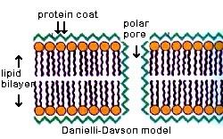  Fluid Mosaic Model A model conceived by S.J. Singer and Garth Nicolson in 1972 to describe the structural features of biological membranes. The plasma membrane is described to be fluid because of its hydrophobicintegralcomponents such as lipids and membrane proteins that move laterally or sideways throughout the membrane. That means the membrane is not solid, but more like a 'fluid'. The membrane is depicted as mosaic because like a mosaic that is made up of many different parts the plasma membrane is composed of different kinds of macromolecules, such as integral proteins, peripheral proteins, glycoproteins, phospholipids, glycolipids, and in some cases cholesterol, lipoproteins. According to the model, the plasma membrane is a lipid bilayer (interspersed with proteins). It is so because of its phospholipidcomponent that can fold in itself creating a double layer - or bilayer - when placed in a polar surrounding, like water. This structural feature of the membrane is essential to its functions, such as cellular transport and cell recognition.  Phospholipids The main constituent of the membrane. The hydrophilic heads of the molecule face the outside and inside of the cell while the hydrophobic tails face each other in the middle of the membrane. Cholesterol This lipid with the four fused rings structure acts to stiffen the membrane and control its fluidity. Proteins Membrane proteins have many different functions. Protein molecules in their tertiary three dimensional form can act as channels, carriers and pumps, cell recognition proteins, receptors for hormones or other signal molecules, and enzymes. The function of membrane proteins will be the topic of other lessons in this unit. Carbohydrates Carbohydrate chains are found attached to membrane lipids and proteins on the cells outer surface. These molecules called, glycolipids or glycoproteins, are responsible for giving each cell a unique identity. Our bodies recognize these cells as our own. Transplanted organs and incompatible blood types have different sugar chains than our own and are attacked by our immune systems causing rejection.
|
| The Following User Says Thank You to AFRMS For This Useful Post: | ||
dr.atifrana (Monday, June 08, 2009) | ||
|
#109
Friday, June 05, 2009
|
|||
|
|||
|
Integral Membrane Proteins
Many of the proteins associated with the plasma membrane are tightly bound to it.
Some transmembrane proteins that span the bilayer several times form a hydrophilic channel through which certain ions and molecules can enter (or leave) the cell.  Peripheral Membrane Proteins These are more loosely associated with the membrane. They are usually attached noncovalently to the protruding portions of integral membrane proteins. Membrane proteins are often restricted in their movements. A lipid bilayer is really a film of oil. Thus we might expect that structures immersed in it would be relatively free to float about. For some membrane proteins, this is the case. For others, however, their mobility is limited:
 Membrane Transport of Small Molecules Because of the hydrophobic interior of the lipid bilayer, polar molecules cannot enter the cell. However, cells devised means of transferring small polar molecules. Transport proteins, each specialized for a certain molecule, can transport polar molecules across the membrane. There are several types of membrane transport proteins. Uniports simply move solutes from one side to another. Cotransport systems work by simultaneously sending two solutes across the lipid bilayer. There are two types of cotransport systems - symport, in which the solutes are sent in the same direction, or antiport, in which they are sent in opposite directions. These transport proteins work passively, meaning that the cell doesn't have to expend energy sending the solute in or out. This is dependent on the solute moving in its natural direction - i.e. moving from more concentrated solution to less concentrated, or from positive to negative. Some specific examples of transport membranes are channel proteins, which allow solutes to cross if they are the correct size and charge. Carrier proteins bind to the solute and lead it through the bilayer. These are examples of passive transport. To move a solute against their natural direction - for example higher concentration to lower concentration, energy (ATP) is needed to pump the solute in or out. An example of active transport is the sodium-potassium pump, which in conjunction with the potassium leak channel allows the cell the control it's membrane potential. The sodium-potassium-ATPase, which uses the energy of ATP hydrolysis, pump pumps sodium out and potassium in, which creates a high concentration of potassium inside the cell, and a low concentration outside. The reverse applies to the sodium. The potassium leak channel allows the potassium to leak out (so to even out the concentrations), which gives the cell and negative charge on the inside. Membrane Transport of Macromolecules Most cells use exocytosis and endocytosis to secrete and ingest macromolecules, respectively. In exocytosis the contents of special vesicles are released when the vesicle fuses with the cell membrane. In endocytosis the membrane depresses and pinches off, enclosing the molecule. Two different sizes are formed - pinocytotic (small) and phagocytic (large). In receptor-mediated endocytosis, coated pits and vesicles bind to specific receptors on the cell surface, allowing the cell to select what molecules to take and what to reject. Membrane Receptors The cell membrane is pocketed with receptors and antigens. Molecules targeted toward that specific cell will bind with the cell surface receptor, which binds the signaling molecule and sends a signal that alters the behavior of the target cell. Antigens are used to tell the cell whether foreign materials are present. If any foreign materials are detected the immune system will mobilize its killer T-cells to destroy the foreign cell. Functions of cell membrane: 1). The cell membrane surrounds the cytoplasm of a cell and, in animal cells, physically separates the intracellular components from the extracellular environment. 2).The cell membrane also plays a role in anchoring the cytoskeleton to provide shape to the cell, and in attaching to the extracellular matrix to help group cells together in the formation of tissues. 3).The barrier is selectively permeable and able to regulate what enters and exits the cell, thus facilitating the transport of materials needed for survival. 4).Specific proteins embedded in the cell membrane can act as molecular signals that allow cells to communicate with each other. Protein receptors are found ubiquitously and function to receive signals from both the environment and other cells. 5). Other proteins on the surface of the cell membrane serve as "markers" that identify a cell to other cells. The interaction of these markers with their respective receptors forms the basis of cell-cell interaction in the immune system.
|
| The Following User Says Thank You to AFRMS For This Useful Post: | ||
dr.atifrana (Monday, June 08, 2009) | ||
|
#110
Friday, June 05, 2009
|
|||
|
|||
|
Lysosomes
Lysosomes are the cells' garbage disposal system. Lysosomes are membrane bound vesicles containing various hydrolytic enzymes necessary for digesting certain material in a cell. Lysosomes contain an ionic pump, which maintains a highly acidic pH They digest excess or worn-out organelles, food particles, and engulfed viruses or bacteria. The membrane surrounding a Lysosomes allows the digestive enzymes to work at the 4.5 pH they require. Lysosomes fuse with vacuoles and dispense their enzymes into the vacuoles, digesting their contents. They are created by the addition of hydrolytic enzymes to early endosomes from the Golgi apparatus. The name Lysosomes derives from the Greek words lysis, which means dissolution or destruction, and soma, which means body. They are frequently nicknamed "suicide-bags" or "suicide-sacs" by cell biologists due to their role in autolysis. 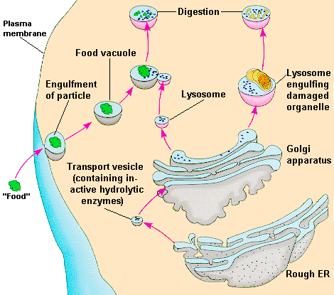 Structure Lysosomes sizes, microscopic appearances, and other properties vary among different cell types and circumstances owing, in part, to differences in their functions and states. Typical Lysosomes are roughly spherical or elongate bodies with largest dimensions of 0.11 micrometer or greater; tens to hundreds are present in a single cell. Each Lysosomes is bounded by a membrane and contains several dozen different species of digestive enzymes, each of which can sever particular chemical bonds found in natural materials. Most lysosomal enzymes function best in an acid environment. This acidification is accomplished by a proton pump, built into the membrane surrounding the Lysosomes, which affects the transport of hydrogen ions into the Lysosomes. Discovery Lysosomes were discovered by the Belgian cytologist Christian de Duve in 1955.He discovered them when he homogenized some animal cells into various components by running them through an ultracentrifuge. After a few days the level of a few enzymes rose significantly implying that they were segregated in the cell and had not attacked any part of the cell before they were broken down. Lysosomes have 2 main roles:
In heterophagy, the cell takes up particles or molecules by the process of endocytosis, engulfing them in membrane-bounded vesicles or vacuoles that are formed at the cell surface. The endocytosed material enters Lysosomes via intermediate membrane-bounded compartments known as endosomes. In higher animals, heterophagy is most prominently used by leukocytes and macrophages. These specialized cells endocytose invasive microorganisms and use endocytosis in clearing debris and disposing of dead or senescent cells. Autophagy In autophagy, cells segregate regions of their own cytoplasm within compartments that come to be bounded by single membranes and to receive lysosomal enzymes. Autophagic lysosomes take part in the remodeling of cells as part of the processes of development and during stressful circumstances. They also participate, along with nonlysosomal enzymes and heterophagic Lysosomes, in normal turnover of the body's constituentsthe balanced synthesis and destruction through which new molecules replace most molecules of most cells. Enzymes To accomplish the tasks associated with digestion, the lysosomes use some 40 different types of hydrolytic enzymes, all of which are manufactured in the endoplasmic reticulum and modified in the Golgi apparatus. Some important enzymes found within lysosomes include:
Clinical relevance There are a number of lysosomal storage diseases that are caused by the malfunction of the lysosomes or one of their digestive proteins, e.g., Tay-Sachs disease, or Pompe's disease. These are caused by a defective or missing digestive protein, which leads to the accumulation of substrates within the cell, impairing metabolism. Genetic defects in lysosomal enzymes and related proteins are known to be associated with a large number of rare disorders in humans and animals (such as Tay-Sachs disease and Niemann-Pick disease type C). Defective lysosomal function leads to storage of particular classes of molecules that cannot be degraded and, in long-lived cells such as neurons, to complex pathogenic cascades with widespread impact on endosomal-lysosomal function, membrane trafficking, and signal transduction. Such disorders are most often fatal. Lysosomes or prelysosomal structures also have been adopted as intracellular homes by certain pathogenic microorganisms that avoid or survive the attacks of the lysosomal system. Some strains of viruses, and toxins such as the one responsible for diphtheria, may use endosomes as their route of entry into the cell, penetrating through the endosomal membrane into the surrounding cytoplasm. Functions Lysosomes are used for the digestion of macromolecules from phagocytosis (ingestion of other dying cells or larger extracellular material, like foreign invading microbes), endocytosis (where receptor proteins are recycled from the cell surface), and autophagy (wherein old or unneeded organelles or proteins, or microbes that have invaded the cytoplasm are delivered to the lysosome). Autophagy may also lead to autophagic cell death, a form of programmed self-destruction, or autolysis, of the cell, which means that the cell is digesting itself. Other functions include digesting foreign bacteria (or other forms of waste) that invade a cell and helping repair damage to the plasma membrane by serving as a membrane patch, sealing the wound. In the past, lysosomes were thought to kill cells that were no longer wanted, such as those in the tails of tadpoles or in the web from the fingers of a 3- to 6-month-old fetus. While lysosomes digest some materials in this process, it is actually accomplished through programmed cell death, called apoptosis. 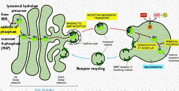 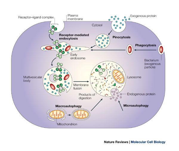 Peroxisomes In 1965, Christian de Duve and his colleagues found other enzymes containing organelle. They called these peroxisomes because they seemed to generate and break down hydrogen peroxide. Peroxisomes, also known as microbodies, and they are self-replicating cells containing oxidative enzymes. They are similar to lysosomes. Their enzymes have two functions; to convert fats to carbohydrates and to detoxify potentially harmful molecules which form in the cell. Peroxisomes, in contrast to lysosomes, are produced only on the smooth ER system. They are found in the cytoplasm of many eukaryotic cells as well as prokaryotic cells, microorganisms, and plant cells. They are very active in yeast, protozoans, kidney cells, and mammailain cells. Peroxisomes are permeable. This allows many small molecules to enter easily. The enzymes of peroxisomes remove hydrogen atoms from small molecules and join them to oxygen creating hydrogen peroxide. They consume up to twenty percent of the oxygen in the liver cell. The peroxisomal enzyme catalase uses this oxygen to convert hydrogen peroxide to water and oxygen neutralizing it. In the liver this method is used to break down molecules of alcohol into substances that can be eliminated from the body. Diseases: The rare fatal genetic disorder Zellweger's syndrome is the result of malformed peroxisomes. This indicates peroxisomes do have an important role in healthy cells.
|
| The Following User Says Thank You to AFRMS For This Useful Post: | ||
dr.atifrana (Friday, June 05, 2009) | ||
 |
«
Previous Thread
|
Next Thread
»
|
|

 Similar Threads
|
||||
| Thread | Thread Starter | Forum | Replies | Last Post |
| Very Important : How to Prepare Study Notes | Shaa-Baaz | Tips and Experience Sharing | 5 | Sunday, May 21, 2017 08:30 PM |
| Effective Study Skills | Sureshlasi | Tips and Experience Sharing | 1 | Friday, November 16, 2007 09:28 AM |
| Regarding Notes | Anonymous84 | Tips and Experience Sharing | 1 | Wednesday, August 15, 2007 06:56 PM |

