CSS Forums
Thursday, April 25, 2024
09:02 AM (GMT +5)
09:02 AM (GMT +5)


|
#11
 Tuesday, April 21, 2009
|
|||
|
|||
|
lectures in biology,covers many topics of zoology.i hope you find this link useful.
http://www.jochemnet.de/fiu/BSC1011/BSC1011_LN.html regards. 
|


| The Following 2 Users Say Thank You to AFRMS For This Useful Post: | ||
dr.taqi abass (Saturday, November 19, 2011), Shali (Thursday, October 08, 2009) | ||
|
#12
Tuesday, April 21, 2009
|
|||
|
|||
|
Vertebrates
This is a link to 60 slide power point presentation about vertebrates. http://www.scribd.com/doc/7748247/Vertebrates regards
|
| The Following 3 Users Say Thank You to AFRMS For This Useful Post: | ||
dr.taqi abass (Saturday, November 19, 2011), Viceroy (Tuesday, April 21, 2009), Shali (Thursday, October 08, 2009) | ||
|
#13
Thursday, April 23, 2009
|
|||
|
|||
|
Protozoans
Protozoa is a subkingdom (formerly a phylum) comprised of organisms with eucaryotic cells that have many of the intracellular components characteristic of higher forms of life. Such other organisms as bacteria do not have this nucleus and are referred to as prokaryotes. Protozoa also have some form of active locomotion, which is a distinguishing feature in classifying them. Even though there are about 50,000 species of protozoa, relatively few are able to cause disease in humans. Protozoal diseases used to be limited to tropical, subtropical, and underdeveloped nations. Now, however, they are becoming a worldwide concern. Protozoa are generally free-living, but some exist as parasites or in a commensal relationship with another organism. The protozoa that are pathogenic parasites are of major interest because they cause such diseases in humans as malaria, trypanosomiasis, toxoplasmosis, and dysentery. Protozoa generally exist in two basic forms: the active, growing form called the "trophozoite;" and the dormant, resistant form called the "cyst." The trophozoite form proliferates tissues, causing damage that results in clinical disease. The cyst is able to survive in an external environment and is usually the form that is transmitted from host to host. Some protozoa go through an intermediate stage in blood-sucking insects. Protozoology is the scientific study of protozoa. Classification of the organism is divided into seven phyla: Sarcomastigophora, Labyrinthomorpha, Apicoplexa, Microspora, Acetospora, Myxozoa, and Aliophora. In 1985, an extensive classification scheme was proposed for protozoa that included various phyla, subphyla, classes, etc. The four groups of protozoa that are mainly responsible for human disease include the following: sarcodina, ciliophora, mastigophora, and sporozoa -- all grouped according to their form of locomotion.
  
|
| The Following 6 Users Say Thank You to AFRMS For This Useful Post: | ||
dr.taqi abass (Saturday, November 19, 2011), farheen79 (Monday, August 26, 2013), Viceroy (Thursday, April 23, 2009), Nazento (Sunday, October 27, 2013), saad sarwar (Tuesday, September 10, 2013), Shali (Thursday, October 08, 2009) | ||
|
#14
Thursday, April 23, 2009
|
|||
|
|||
|
|
| The Following 5 Users Say Thank You to AFRMS For This Useful Post: | ||
dr.taqi abass (Saturday, November 19, 2011), Viceroy (Thursday, April 23, 2009), Muhammad T S Awan (Monday, October 19, 2009), saad sarwar (Tuesday, September 10, 2013), Shali (Thursday, October 08, 2009) | ||
|
#15
Friday, April 24, 2009
|
|||
|
|||
|
Reproduction in Protozoa
Asexual Reproduction Asexual reproduction is the most common means of replication by protozoans. The ability to undergo a sexual phase is confined to the ciliates, the apicomplexans, and restricted taxa among the flagellates and sarcodines. Moreover, sexual reproduction does not always result in an immediate increase in numbers but may simply be a means of exchanging important genetic materialbetween individuals of the same species (conjugation). Free-living protozoans normally only resort to sexual reproduction when environmental conditions become adverse, because this mode of reproduction enhances the fitness of the population and increases the chance of mutation. When food and other conditions are favourable, asexual reproduction is practiced. Asexual reproduction in free-living species usually involves nuclear division and the division of the cell into two identical daughter cells of equal size by binary fission. In parasitic protozoa and some free-living species, multiple fission, resulting in the production of many offspring that may not resemble the parent cell, is normal. During the cycle of growth and division, the protozoan undergoes a series of identifiable phases: a division phase, a growth phase during which the cell increases substantially in size, a phase of DNA synthesis, and a phase of preparation for division, which extends from the end of DNA synthesis until the initiation of division. The division of the cytoplasm is preceded by the division of the nucleus or nuclei. The plane of division in protozoan cells varies among the different groups and is of taxonomic significance. The flagellates normally divide in a longitudinal plane. The usual process starts at the front end with the division of the flagella and the associated structures; simultaneously, the nucleus divides. The cytoplasm then splits from front to back into two identical daughter cells. The ciliates normally divide in an equatorial, or transverse, plane, thereby maintaining the correct number of ciliary rows, or kineties. The cell mouth and any specialized cilia around it are replicated in different ways among the various ciliate groups, depending on the complexity of the cytostome. The replication of the cytostome precedes the division of the cytoplasm. Some ciliates (e.g., Colpoda) divide within thin-walled reproductive cysts into two daughter ciliates, each of which then divides so that the cyst contains four progeny, which are released when the cyst wall ruptures. The sedentary suctorians do not reproduce by binary fission because the production of an identical, nonswimming offspring would rapidly lead to overcrowding. They instead produce single ciliated offspring called swarmers by a process called budding. Budding can occur endogenously, in which the bud forms within the parent and is ejected when mature, or exogenously, in which the swarmer is formed outside the parent. The swarmers swim away from the parent, settle on a substrate, lose their cilia, and develop feeding tentacles and an attaching stalk. Naked amoebas (rhizopods) have no fixed plane of division but simply round up and divide into two basically equal halves. The testate amoebas (also rhizopods), which live in single-chambered shells, or tests, exude the daughter from the aperture of the shell. In species that have a shell formed from silica plates, the daughter contains the plates used to produce the shell but remains attached to the mother cell until the shell is fully formed, when the final severing of the cytoplasm between the individuals occurs. Some of the testate amoebas live inside proteinaceous shells. There, too, the new shell is secreted before binary fission is completed. The foraminiferan and radiolarian sarcodines have evolved multiple fission. Both produce many flagellated swarmers, or zoospores. The common planktonic foraminiferan Globigerinoides sacculifer, for example, can produce 30,000 swarmers at one time. Each swarmer is about 5 micrometres (0.005 millimetre) long. In planktonic species the parent usually loses buoyancy and sinks by shedding spines and withdrawing the complicated pseudopodial network into the shell. The swarmers are produced in deep water and migrate upward as they mature. Each secretes a shell around itself, which is added to as the organism grows. The foraminiferans are unusual among free-living protozoans in that a sexual phase is a regular part of the life cycle, alternating with an asexual phase. During the life cycle two types of swarmer are produced. One type, zoospores, have half the number of chromosomes of the parent (i.e., they are haploid); they grow until they become mature adults and can produce and release large numbers of gametic swarmers. These gametes are identical (isogamous) but are comparable to the eggs and sperm of higher organisms. The gametic swarmers fuse in pairs, thus restoring the full complement of chromosomes (i.e., they are diploid), and each individual grows, matures, and ultimately produces haploid zoospores. Sexual reproduction among the flagellates is not widespread and can involve identical gametes (isogamy) or distinct male and female gametes (anisogamy). The female gametes are larger and are stationary, whereas the male gametes are smaller, produced in larger numbers, and motile. Sexual Reproduction Sexual reproduction among the ciliated protozoans takes the form of conjugation. The process does not result in an increase in numbers, but is a simple exchange of genetic material between two individual cells. Conjugation occurs only between compatible mating strains within a species, and each species may contain many mating strains. Before conjugation occurs, special chemical signals, called gamones, are released by some ciliates. The gamones cause compatible mating strains to undergo processes that facilitate conjugation. In other ciliates, such as Paramecium, gamones are bound to the cell surface and elicit their responses when the ciliates make physical contact. During conjugation, two ciliates line up side by side. The macronucleus, which plays no part in the process, disintegrates. A series of nuclear divisions of the micronuclei in each ciliate then ensues, including a meiosis, during which a number of haploid micronuclei are produced in both cells. All but one of these haploid micronuclei disintegrate. The remaining haploid micronucleus in each cell then divides through mitosis into two haploid nuclei (gamete nuclei). A bridge of cytoplasm forms between the two ciliates, and one gametic nucleus from each cell passes into the other cell. The two gametic nuclei in each cell unite, thus restoring the diploid number of chromosomes. The micronucleus undergoes two mitotic divisions to produce four micronuclei; two of these will form the new micronuclei of the cell and two are destined to become the macronucleus. Following the process of conjugation, normal binary fission proceeds. The number of macronuclei and micronuclei formed is dependent on the species and remains the same as the original number. When no suitable mating partner is available, ciliates may undergo a form of conjugation called autogamy, in which all of the nuclear processes described above occur. But, because only one individual is involved, there is no exchange of gametic nuclei; instead, the two gametic nuclei within the cell unite to form the restored micronucleus. Specialized sedentary suctorian ciliates practice a modified form of conjugation. The conjugating individuals differ in appearance. The macroconjugants resemble the normal feeding individuals, and the microconjugants resemble the swarmers, although smaller. When a microconjugant locates a macroconjugant, it enters and fuses with it. This is quite different from the temporary association between two cells that occurs in most ciliates. As is common with other parasitic organisms, parasitic protozoans face the problem of how to disperse from one host to another. In order to increase the probability of finding more hosts, most parasitic protozoa reproduce in high numbers. A representative life cycle of a parasitic protozoan can be found in members of the parasitic phylum Apicomplexa. These protozoans have a complex life cycle that involves a series of stages characterized by episodes of asexual multiple division called schizogony. In the parasite Plasmodium, for example, this phase of the life cycle occurs in the liver and red blood cells of humans. The parasite (sporozoite) enters the hosts cells and grows while feeding on the cell contents. It then undergoes a multiple asexual division (schizogony) into many individuals (merozoites). The hosts cell wall ruptures, permitting each individual to invade a new red blood cell and repeat the process. In certain merozoites a sexual cycle is eventually initiated inside the red blood cell, and male and female gametes are produced. The male gametes (microgametocytes) are small, while the female gametes (macrogametocytes) are larger. The life cycle continues if the gametocytes are taken up by a feeding female mosquito of the genus Anopheles. Only the gametocytes can infect the mosquito. Inside the mosquitos gut the haploid gametes fuse to form a diploid zygote, which then undergoes sporogony, a process of multiple divisions in which many sporozoites are produced. The sporozoites migrate to the salivary glands of the insect and are injected into a new host when the mosquito next feeds. They are carried by the blood to the liver, where they undergo their first schizogony inside liver cells, thereafter invading the red blood cells for repeated cycles of schizogony. The parasitic flagellates reproduce entirely by asexual means and do not appear to have a sexual phase in their life cycles. There is, however, evidence of genetic exchange between certain subspecies of Trypanosoma brucei, although the process by which this occurs is not known. 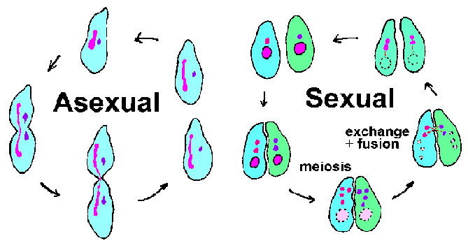 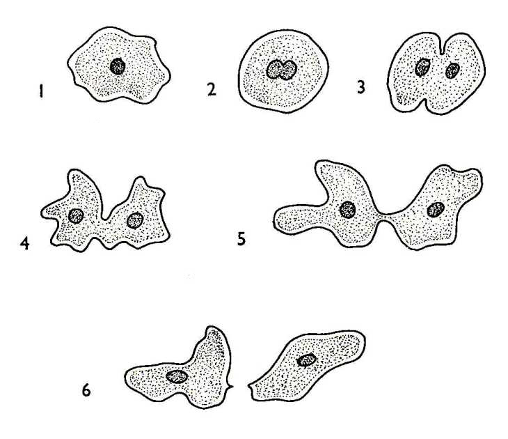 Last edited by Viceroy; Friday, April 24, 2009 at 08:00 AM. Reason: Formatting
|
| The Following 3 Users Say Thank You to AFRMS For This Useful Post: | ||
dr.taqi abass (Saturday, November 19, 2011), Viceroy (Friday, April 24, 2009), Shali (Thursday, October 08, 2009) | ||
|
#16
Friday, April 24, 2009
|
|||
|
|||
|
|
|
#17
Friday, April 24, 2009
|
|||
|
|||
|
The Phylum Porifera
Etymology:- From the Latin porus for pore and Ferre to bear, hence an animal with with pores. Characteristics of Porifera:- 1)No definite symmetry. 2)Body multicellular, few tissues, no organs. 3)Cells and tissues surround a water filled space but there is no true body cavity. 4)All are sessile, (live attached to something as an adult). 5)Reproduce sexually or asexually, sexual reproduction can be either gonochoristic or hermaphroditic. 6)Has no nervous system. 7)Has a distinct larval stage which is planktonic. 8)Lives in aquatic environments, mostly marine. 9)All are filter feeders. 10)Often have a skeleton of spicules. Sponges are one of the better known groups of invertebrates, due to their usefulness in the bath many people who care nothing for invertebrates at least know name and may even have seen a sponges skeleton on sale in a shop. One of the more amazing things about sponges is there ability to suffer damage. Because the cells are not linked in a tissue it is possible for them to be separated an then come together again. Some species such as the freshwater sponge Ephydatia fluviatilis can be pushed through a sieve, then if given time the individual cells will come together again and make a new sponge. A sponge is a simple organism that is easy to describe. A sponge is a sedentary, filter-feeding metazoan which has a single layer of flagellated cells that drive a unidirectional current of water through its body. such a brief description though does not do them justice. Sponges are an ancient and highly successful group of animals. In the Palaeozoic they are believed to have comprised more than half the biomass in marine reefs. They have been living in the waters of the world for more than 600 million years, and can now be found in all marine and many freshwater habitats. Sponges occur in rivers and streams, from rock pools to the deep ocean depths, from frozen arctic seas to the warm tropical seas. They are perhaps at their most beautiful in tropical marine seas. There are about 10,000 known species and though their basic organisation is pretty simple and remains fairly constant throughout the all species they do manage to show a great variety of forms. Anatomy The body of a sponge is a collection of a few different types of cells loosely arranged in a gelatinous matrix called a 'mesohyl', mesoglea or mesenchyme. This mesohyl is the connective tissue of a sponge body and it is supported by the skeletal elements. The skeletal elements of sponges are variable and important in taxonomy. Throughout this body run canals through which water flows, there is considerable variation in the complexity of these canals. The canals have openings to the outside which are called pores, where the water enters the sponge system these pores are usually small and are called 'ostia' and where the water leaves the sponge system the pores are larger, often singular and are called 'oscula' (singular osculum). Many if not most of these canals are lined with special flagellated cells called 'choanocytes'. These choanocytes keep the water flowing through the canals in the correct direction by beating their flagellum, they are also important in trapping food items. There are three main types of canal system in sponges. The simplest form is Asconoid, here the canals run straight through the sponge body and all the choanocytes line the central large space called the 'spongocoel'. The water enters the ostia, is drawn through to the spongocoel and leaves through a single large osculum. Asconoid sponges have cylindrical hollow bodies and tend to grow in groups attached to some object or other in relatively shallow seas. Slightly more complicated are Syconoid sponges, externally they are fairly similar to asconoid sponges except that their body wall is thicker. The canals are branched however and do not allow the water to flow straight through in to the spongocoel. Instead the water flows a twisted route through a number of canals some of which are lined with choanocytes before being expelled into the spongocoel and out through the osculum. The spongocoel is not lined with choanocytes only the canals. Syconoid sponges go through a asconoid stage in their development suggesting that they evolved from some ancestral asconoid. Syconoid sponges do not normally form groups as do asconoid sponges. Most modern sponge species are Leuconoid. In leuconoid sponges the canal system is more complicated again with the canals being longer and more branched, they lead to special chambers whose walls are lined by choanocytes, there are no choanocytes in the canals. There is no real spongocoel just a central exit canal leading to the osculum. Leuconoid sponges tend to live in large groups with each individual sponge having its own osculum, however the borders between individual sponges are often hard to define and the sponge may act more like a large communal organism. Sponges are built up from relatively few cell types, the main ones being choanocytes, pinacocytes, amoebocytes and lophocytes. Choanocytes are vase shaped cells with a collar of fine fibrils connected by microvilli. this is a filter which strains out the smallest food items from the water such as individual bacteria. Extending from the centre of this collar is the single flagellum whose beating drives the water currents that keep the sponge alive and healthy. Pinacocytes, these form much of the epidermis of sponges and are as close as a sponge gets to having a tissue. Generally they cover the exterior and some interior surfaces. They can change their size (they are contractile) and can therefore change the size of the openings of the ostia thus controlling the flow of water through the sponge. Pinacocytes are also implicated in the absorption into the sponge of larger food items. Amoebocytes come in several forms, they are alike in that they are mobile and move around within the sponge body. Archaeocytes are the basis of some asexual reproductive gemmules. If an amoebocyte secretes the spongin fibres of the skeleton they are called a spongioblast, if it secretes spicules it is called a scleroblast and if it is star shaped and secrete collagenous fibrils then it is called a collencyte. Lophocytes are a type of amoebocyte, they are the most motile of the sponge cells moving around relatively freely within the mesohyl where they are important in the secretion of fibrils. Sponges have skeletons, if it were not so they would be just blobs. There are two main components of a sponge skeleton, a protein called spongin which forms a tough fibrous network throughout the sponge and normally works in conjunction with the spicules. Spicules are non-living aggregates of a chemical nature, secreted and made from either silica or calcium carbonate as calcite or aragonite. These spicules are important in the classification of sponges, thus we can say that. The Calcarea sponges have spicules of calcium carbonate that have 1,3 or 4 rays, a a skeleton that involves a single large lump of calcium carbonate rather than spicules. The Demospongiae have their spicules made from silica and they have 1,2, or 4 rays. The Sclerospongiae have a compound skeleton of spicules of silica that is restricted to thin layer of living sponge supported on a large basal layer of calcium carbonate. The Hexactinellida or 'Glass sponges' have spicules made from silica that are 6 rayed. Individual spicules can be arranged loosely within the spongin or interlocking and fused together, siliceous spicules come in two sizes called megascleres and microscleres. Ecology All sponges are filter feeders on small to extremely small particles and most are sedentary or immobile as adults, i.e they spend their adult lives fixed to a substrate. The reproductive ecology of most sponges has never been studied so the following generalisation is based on the few species that are reasonably well known and should not be taken as the last word in sponge reproductive ecology. Sponges are generally hermaphroditic, however they are only one gender at a time, being either male or female or neuter, some species such as Halichondria moorei change colour when they change sexes though most do not. Sponges have no permanent gonads, instead a number of areas of the sponge will during the reproductive period become differentiated (changed) to produce either sperm or ova (eggs). Sperm is released into the canals and is then pumped out of the sponge through the osculum where it is likely to be drawn into the canal system of another sponge. Here incoming sperm of the same species are trapped by the choanocytes which then loose their flagellum and collar and migrate through the mesohyl to the ovocyte, a cell generating ova, where the sperm are transferred to the ova, assuming this is a sponge in its female form. Sperm release can be an individual act as in Verongia archeri or it can be a co-ordinated affair with many sponges in an area releasing their sperm simultaneously as in Neofibularia nolitangere. The fertilised ova are retained within the adult sponge until some unknown signal indicates it is time for their release. They are then set free into the surrounding waters. Once an adult begins expelling its larvae it continues to do so for some time, thus Microciona coccinea releases 4 or 5 larvae per minute for 3 to 4 days. Larval sponges are small 50 microns to 5 millimetres in diametre. all known sponge larvae are ciliated though the cilia may be longer, shorter or absent from different parts of their surface. After release they swim or crawl for a period of time before settling down to begin life as a new miniature sponge. Swimming species tend to have a crawling phase immediately before settling down. This free living stage may last as long as 18 to 20 days in Polymastia spp. or be as short as 4 to 6 hours in genera such as Ophlitaspongia. Larval sponges are not complicated organisms, and there is much variation between species however many species have a positive phototaxis when they first leave their parents body which switches to a negative one before they enter the presettling stage. Some species have been shown to have a preliminary negative geotaxis while most species have shown a preference for surfaces with an algal or bacterial film. As with the larval stage so the time taken for the larvae to reorganise itself into and functioning sponge varies between species so that Microciona spp. are up and running within two days while Polymastia spp. can take as long as 7 days to get themselves sorted. Sponges also reproduce asexually by releasing fragments of themselves, or special groups of cells called gemmules. These gemmules, at least in freshwater species such as Ephydatia fluviatilis have protective coat of spongin and have particular environmental conditions they need to have met before they germinate.  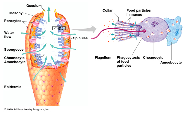 Last edited by AFRMS; Friday, April 24, 2009 at 03:19 PM. Reason: Formatting
|
| The Following 2 Users Say Thank You to AFRMS For This Useful Post: | ||
Imadafridi (Tuesday, May 12, 2009), Shali (Thursday, October 08, 2009) | ||
|
#18
Friday, April 24, 2009
|
|||
|
|||
|
Canal System in Porifera.
There are three types of canal systems, in the following drawings the arrows show the direction of water flow: Asconoids have the simplest, see the drawing on the left. These sponges are small and tube-shaped. The water enters through tiny ostia into one large internal cavity called a spongocoel, and is expelled through one large osculum. This type of canal system is found in the Calcarea Class . Synconoids, like asconiods, have a single, large osculum, but their body is thicker. The drawing below left shows two synconoid systems - a simple type on the left and a more complex type on the right. The water enters through numerous small ostia, and passes through incurrent canals before reaching the large central cavity. This system is found in Calcarea and Hexactinellida . Leuconoids have the most complex structure . They have many small ostia. The ostia lead to numerous incurrent canals, but there is no large central cavity. Asconoid Type    Three Types of canal system 
|
| The Following 2 Users Say Thank You to AFRMS For This Useful Post: | ||
dr.taqi abass (Saturday, November 19, 2011), Shali (Thursday, October 08, 2009) | ||
|
#19
Friday, April 24, 2009
|
|||
|
|||
|
Phylum Porifera
The link mentioned here cantains very good presentation about porifera,structure i.e skeleton,canal system ,reproduction.... with very good figures. Click Here. Regards
|
| The Following User Says Thank You to AFRMS For This Useful Post: | ||
Shali (Thursday, October 08, 2009) | ||
|
#20
Saturday, April 25, 2009
|
|||
|
|||
|
Skeleton in Protozoa , a very good and interesting presentation on protozoa skeleton,do check it.
Click this. regards
|
 |
«
Previous Thread
|
Next Thread
»
|
|

 Similar Threads
|
||||
| Thread | Thread Starter | Forum | Replies | Last Post |
| Very Important : How to Prepare Study Notes | Shaa-Baaz | Tips and Experience Sharing | 5 | Sunday, May 21, 2017 08:30 PM |
| Effective Study Skills | Sureshlasi | Tips and Experience Sharing | 1 | Friday, November 16, 2007 09:28 AM |
| Regarding Notes | Anonymous84 | Tips and Experience Sharing | 1 | Wednesday, August 15, 2007 06:56 PM |

