CSS Forums
Friday, May 03, 2024
01:57 AM (GMT +5)
01:57 AM (GMT +5)
|
|||||||


 |
Share Thread:
 Facebook
 Twitter
 Google+
|
|
|
LinkBack | Thread Tools | Search this Thread |
|
#1
 Thursday, September 29, 2011
|
||||
|
||||
|
THE DISCOVERY OF CELLS
Antony Van Leeuwenhoek built this simple microscope over three hundred years ago. He used it to discover single celled organisms swimming around in pond water. What Leeuwenhoek imagined, was that the tiny organisms he observed were actually little animals. He called them his “cavorting wee beasties”. He even recorded their means of reproducing. They duplicated their structures and then pulled apart to create two identical individuals. This kind of reproduction is quite different than having babies or growing a new plant from a seed. In these microscopic organisms one individual duplicates itself becoming identical twin sisters.Around the same time Leeuwenhoek was observing his “wee beasties,” a British scientist, Robert Hook, was turning his more elaborate microscope onto a shaving of cork, just to see if it had a hidden structure of some kind. He saw that the cork was made up of thousands of little compartments. They reminded him of the prison cells where prisoners were held. Thus the term — cell. CELL STRUCTURES  Around a hundred years ago microscopes showed that all cells have a common structure. All have an outer membrane that holds the cell together, a membrane that allows some substances to pass, but excludes others. The cells of plants, animals and “protists” as Leeuwenhoek’s wee beasties came to be called, all contain a nucleus. It was soon realized that this structure somehow controlled the cell’s activities. The nucleus was seen to duplicate just before cell division, suggesting that it carried hereditary information from one generation of cells to the next. In cells undergoing division the nucleus broke down and its internal structures became visible -- chromosomes Each species has a characteristic chromosome number. Humans have 23 pairs of chromosomes in each of their body cells. Outside of the nucleus is a soupy fluid containing many small bodies, a cell’s organs, and appropriately called organelles. In a cell that has been squeezed by the cover glass to the point of rupture, a bubble forms. In this bubble you can clearly see the tiny cell organelles called mitochondria. Similar organelles were eventually found in all cells, both plant, animal and protist. Mitochondria were eventually isolated and their function was discovered. They are the bodies where sugars and other food molecules are metabolized, with the help of oxygen, to supply energy for cell use – the process of cellular respiration. Plants and green protists have an additional cell organelle clearly visible: rounded green bodies called chloroplasts. These green bodies are where photosynthesis occurs. In the process of photosynthesis chloroplasts use the energy in light to convert carbon dioxide from the air into sugars and other organic compounds needed by the plant Cells - Structure and Function Important Events in the Discovery of Cells
Cells are complex and highly organized
   Prokaryotes Prokaryotes
Features of Prokaryotic Cells 
Properties of Eukaryotic Cells  A Typical Plant Cell  A Typical Animal Cell
 CELL FUNCTION AND STRUCTURE
__________________
"Wa tu izzu man-ta shaa, wa tu zillu man-ta shaa" 
|


| The Following 5 Users Say Thank You to SADIA SHAFIQ For This Useful Post: | ||
Arain007 (Friday, September 30, 2011), mjkhan (Saturday, October 01, 2011), siraj narejo (Friday, September 30, 2011), SYEDA SABAHAT (Thursday, September 29, 2011), Taimoor Gondal (Sunday, October 02, 2011) | ||
|
#2
Saturday, October 01, 2011
|
||||
|
||||
|
Muscle cell
Muscle is a contractiletissue of animals and is derived from the mesodermal layer of embryonic germ cells. Muscle cells contain contractile filaments that move past each other and change the size of the cell. They are classified as skeletal, cardiac, or smooth muscles. Their function is to produce force and cause motion. Muscles can cause either locomotion of the organism itself or movement of internal organs. Cardiac and smooth muscle contraction occurs without conscious thought and is necessary for survival. Examples are the contraction of the heart and peristalsis which pushes food through the digestive system. Voluntary contraction of the skeletal muscles is used to move the body and can be finely controlled. Examples are movements of the eye, or gross movements like the quadriceps muscle of the thigh. There are two broad types of voluntary muscle fibers: slow twitch and fast twitch. Slow twitch fibers contract for long periods of time but with little force while fast twitch fibers contract quickly and powerfully but fatigue very rapidly.  Muscle cells are individual cells that comprise the muscle tissue of the body and execute muscle contraction. There are three types of muscle cells: skeletal, cardiac, and smooth. Each of these types differ in cellular structure, specific function, and location within the body. Together, the three muscle cell types play specific roles in supporting the skeletal structure and posture of the body, assisting in the flow of blood through blood vessels, aiding in digestion, and driving the heartbeat. Skeletal muscle cells are found throughout the body, making up skeletal muscle that is anchored to the bones by ligaments. During development, skeletal muscle cells are made from precursor cells, called myoblasts, which fuse together to form long, cylindrical, mature muscle cells. Each muscle cell contains several nuclei one from each myoblast that is used to make up the cell and fibers that have striations where the myoblasts were fused together. Skeletal muscle cells allow for muscle contraction, and they are responsible for movement and the upright posture of the body. These cells are voluntary muscle cells, meaning they receive signals from the brain to perform contraction. Cardiac muscle cells are found in the walls of the heart. Like skeletal muscle cells, mature cardiac muscle cells have a striated appearance, which is a result of different protein fibers within the cardiac muscle cell. Each cardiac muscle cell has a number of irregular branches, and each branch is connected to branches on neighbor cells by an adhering structure called an intercalated disc. Cardiac muscle cells are highly resistant to fatigue, and their regular contraction allows for beating of the heart, thereby pumping blood out of the heart and into the blood vessels. These cells are said to be involuntary, since they do not rely on conscious signals from the brain to contract. Smooth muscle is structurally distinct from skeletal and cardiac muscle cells. Unlike these cell types, a smooth muscle cell does not have a striated appearance, and instead forms homogeneous bundles. Smooth muscle cells are found in a number of systems throughout the body; for example, they make up a component of veins and arteries, and surround organs in the gastrointestinal tract. A layer of smooth muscle cells surrounds veins and arteries to provide strength and aid in movement of blood through the vessel. Smooth muscle cells also surround the esophagus, stomach, small intestines, and large intestines to aid in digestion and movement of food through the digestive system. Like cardiac muscle cells, contraction of a smooth muscle cell is involuntary and does not require conscious signals from the brain to contract and perform its functions throughout the body. Anatomy of a muscle cell
__________________
"Wa tu izzu man-ta shaa, wa tu zillu man-ta shaa"
|
|
#3
Sunday, October 02, 2011
|
||||
|
||||
|
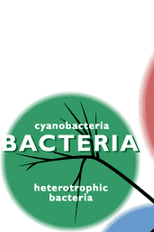
Bacteria
Bacteria are microscopic organisms whose single cells have neither a membrane-enclosed nucleus nor other membrane-enclosed organelles like mitochondria and chloroplasts. Another group of microbes, the archaea, meet these criteria but are so different from the bacteria in other ways that they must have had a long, independent evolutionary history since close to the dawn of life. In fact, there is considerable evidence that you are more closely related to the archaea than they are to the bacteria. Properties of Bacteria
Until recently classification has done on the basis of such traits as:
 Gram-positive bacteria are encased in a plasma membrane covered with a thick wall of peptidoglycan. Gram-negative bacteria are encased in a triple-layer. The outermost layer contains lipopolysaccharide (LPS). The Gram stain is named after the 19th century Danish bacteriologist who developed it. Gram-positive bacteria are encased in a plasma membrane covered with a thick wall of peptidoglycan. Gram-negative bacteria are encased in a triple-layer. The outermost layer contains lipopolysaccharide (LPS). The Gram stain is named after the 19th century Danish bacteriologist who developed it.
More recently, genome sequencing, especially of their 16S ribosomal RNA (rRNA), has provided additional insights into the evolutionary relationships among the bacteria. Firmicutes Comparison of their sequenced genomes reveals that all the Gram-positive rods and cocci as well as the mycoplasmas belong to a single clade that has been named the Firmicutes. Gram-Positive Rods Aerobic Gram-Positive Rods
 The bacteria in this group grow in characteristic colonies. The bacteria in this group grow in characteristic colonies.
Mycoplasmas have the distinction of being the smallest living organisms. They are so small (0.1 µm) that they can be seen only under the electron microscope. Mycoplasmas are obligate parasites; that is, they can live only within the cells of other organisms. They are probably the descendants of Gram-positive bacteria who have lost their peptidoglycan wall as well as much of their genome — now depending on the gene products of their host. The DNA sequences of the complete genomes of seven mycoplasmas have been determined, including
The scientists at The Institute for Genomic Research (now known as the J. Craig Venter Institute — JCVI) who determined the Mycoplasma genitalium sequence followed this work by systematically destroying its genes (by mutating them with insertions) to see which ones are essential to life and which are dispensable. Of the 485 protein-encoding genes, they have concluded that only 381 of them are essential to life. Workers at the JCVI have also succeeded in synthesizing the complete genome of one species of mycoplasma, inserted this into a second species, which converted the second species into the first. Read more about this remarkable achievement. Actinobacteria Most of these Gram-positive organisms grow as thin filaments — like a mold — rather than as single cells. In fact, they were long thought to be fungi and were called actinomycetes. But fungi are eukaryotes and the actinobacteria are not. Actinobacteria dominate the microbial life in soil where they play a major role in the decay of dead organic matter. Many of them have turned out to be the source of valuable antibiotics, including streptomycin, erythromycin, and the tetracyclines. Mycobacteria and Corynebacteria These Gram-positive organisms are closely related to the actinobacteria and often classified with them. They include three important human pathogens:
This large group of bacteria form a clade sharing related rRNA sequences. They are all Gram-negative but come in every shape (rods, cocci, spirilla). They are further subdivided into 5 clades: alpha-, beta-, gamma-, delta-, and epsilon proteobacteria. Alpha (α) Proteobacteria. Some examples:
The largest and most diverse subgroup of the proteobacteria. Some examples <UL>Escherichia coli. The most thoroughly-studied of all creatures (possibly excepting ourselves). Its entire genome has been determined down to the last nucleotide: 4,639,221 base pairs of DNA encoding 4,377 genes. Lives in the human colon, usually harmlessly. However, water or undercooked food contaminated with the O157:H7 strain has caused severe — occasionally fatal — infections. Salmonella enterica. Two major human pathogens:
Pseudomonas aeruginosa. A common inhabitant of soil and water, it can cause serious illness in humans with
Yersinia pestis. This bacillus causes bubonic plague. It is usually transmitted to humans by the bite of an infected flea. As it spreads into the lymph nodes, it causes them to become greatly swollen, hence the name "bubonic" (bubo — swelling of a lymph node) plague. Once in the lungs, however, the bacteria can spread through the air causing the rapidly lethal (2–3 days) "pneumonic" plague. Untreated, ~30% of the cases of bubonic plague are fatal, and the figure for the pneumonic form reaches 100%. The recurrent epidemics of the "black death" in Europe from 1347–1352, which killed off at least one quarter of the population, are thought to have been caused by this organism. DNA sequencing of samples retrieved from the bodies of plague victims of that era confirm this diagnosis.Why are the Gram-negative bacteria encased in two membranes while the Gram-positives have only one? Evolutionary biologist James Lake has proposed that the Gram-negatives arose by one single-membrane bacterial ancestor engulfing another. His analysis of many genes in the various bacterial groups indicate that the most probable ancestors of this possible endosymbiosis were a clostridium and an actinobacterium. Clostridia are the only Gram-positive bacteria that have photosynthetic members and because the photosynthetic apparatus in all photosynthetic Gram-negative bacteria is in the inner membrane, perhaps the actinobacterium was the host and the clostridium the endosymbiont.
__________________
"Wa tu izzu man-ta shaa, wa tu zillu man-ta shaa"
|
|
#4
Sunday, October 02, 2011
|
||||
|
||||
|
Obesity or stem cell research could win Nobel
STOCKHOLM (AP) Two scientists who unlocked some of the mysteries linked to obesity or a professor who figured out how to make stem cells without human embryos could be candidates for the medicine award when the first of the 2011 Nobel Prizes are announced Monday. The prize committees don't give any clues they even keep nominations secret for 50 years but winners usually have won many other awards and distinctions before they are considered for a Nobel. Canadian-born Douglas Coleman and American Jeffrey Friedman have won several prizes for their discovery of leptin, a hormone that regulates food intake and body weight, and could be in the running for the coveted prize worth 10 million kronor ($1.5 million). Last year, Coleman, of the Jackson Laboratory in Bar Harbor, Maine, and Friedman, of Rockefeller University in New York, received the Lasker Award, often seen as a precursor to the Nobel, for having shown that obesity is frequently linked to metabolic disruptions, or the lack of leptin, rather than being a self-induced problem. Japanese Shinya Yamanaka, another potential Nobel candidate, offered the world of regenerative medicine a breakthrough with experiments showing that stem cells can be made from ordinary skin cells. The discovery led to a leap in stem cell research, reducing the need for using human embryos. Yamanaka won the Lasker Award in 2009 and this year shared Israel's Wolf Prize. One out of three Wolf award-winners in chemistry, physics and medicine have also won a Nobel Prize. Yamanaka, of Kyoto University in Japan and the Gladstone Institute of Cardiovascular Disease in San Francisco, could share the prize with British cloning pioneer John Gurdon or Canadian stem cell researcher James Till. Till discovered blood stem sells, which have saved the life of many thousands of leukemia patients. "Gurdon's cloning technique and Yamanaka's stem cells are highly interesting in the field of basic science," wrote science reporter Karin Bojs of Swedish daily Dagens Nyheter, who has stood out as a leading Nobel guesser over the years. "But so far, not a single sick person has been cured with these discoveries. It is therefore possible that Yamanaka and Gurdon get to share the prize with Canadian James Till." Bojs said other possible candidates for the prize are the American-French trio Ronald Evans, Elwood Jensen and Pierre Chambon for their research on nuclear hormone receptors, and American David Julius for his discoveries of the molecular mechanisms by which the skin senses pain, heat and cold. "It will be awarded to fundamental discoveries that leads to an understanding of the human body and, or treatment or prevention of illnesses," said Nobel Prize Committee Secretary Goran Hansson, declining to give away more details. He said there are so many Nobel-worthy achievements in medicine that it can be hard to select a winner. In an unusual leak last year, a Swedish newspaper revealed the jury's selection British test tube baby pioneer Robert Edwards before the announcement. The committee has since applied even stricter rules on keeping their discussions and documents surrounding potential candidates secret. But that doesn't keep people from making predictions. The scientific department of Thomson Reuters, which analyzes high-impact academic papers to make predictions, suggested U.S. scientists Brian Druker, Nicholas Lydon, and Charles Sawyers, could be awarded for work related to Gleevec and Sprycel, drugs that transformed chronic myelogenous leukemia from a fatal cancer into a manageable chronic condition. Its predictions also include Robert Langer and Joseph Vacanti "for their pioneering research in tissue engineering and regenerative medicine," as well as Jacques Miller, Robert Coffman and Timothy Mosmann for their discovery of two types of blood cells and their role in regulating immune responses. The Nobel Prizes date back to 1901 after a will left behind by Swedish dynamite inventor Alfred Nobel, and medicine winners are typically awarded for a specific breakthrough rather than a body of research. The other award categories include physics, chemistry, literature and peace. The economics award isn't technically a Nobel and was established by Sweden's central bank in 1968. The prizes are handed out every year on Dec. 10, on the anniversary of Nobel's death in 1896. http://news.yahoo.com/obesity-stem-cell-research-could-win-nobel-092832956.html;_ylt=AlOqRVjhvHfTTUGQrF9PNhes0NUE;_ ylu=X3oDMTNqcWMzOHZkBG1pdAMEcGtnAzg5MzdhMGE3LTg3MT ktMzYyNC1hZDNiLWJiNDliMGNmOGIwOQRwb3MDMTMEc2VjA2xu X0xhdGVzdE5ld3NfZ2FsBHZlcgM2MWM0MjU4MC1lY2U3LTExZT AtYmY5Yi1iYmUzMGI0ODE5Zjc-;_ylv=3
__________________
"Wa tu izzu man-ta shaa, wa tu zillu man-ta shaa"
|
|
#5
Thursday, October 06, 2011
|
||||
|
||||
|
The word micro-organism is a general term for a (very) small organism, so small that the use of a microscope is required to see details of its structure. The study of micro-organisms (also known as microbes) is called microbiology, and it is increasingly relevant in Biology.
A virus is a very small creature and can not be seen by the naked eye. They can only be seen with the aid of a microscope. Their size is so small that their size can only be measured in micro-meters. The organism which is so small and is measured in micros are known as micro organisms  Main groups of micro-organismsStudy calledSpecialist called viruses (not really living organisms like the rest)virology(virologist)bacteriabacteriology(bacteriologist)fungimycology(mycologist)possibly less important protozoaprotozoology(protozoologist)algaealgology (phycology)algologist (phycologist) Each of these groups includes organisms which can be seen to be useful to man (especially in the context of biotechnology), and others which are harmful (mainly because of diseases - of animals and plants, or spoilage - of stored products, especially food). There are broad similarities in the way that these micro-organisms grow, but there are distinct differences in detail which must be appreciated. From a classification point of view, these micro-organisms are now thought to merit separating from other more familiar living organisms (Plants and Animals), so they have been given Kingdoms of their own: Group of micro-organismKingdomBacteria and Blue-green "algae"MoneraProtozoa and AlgaeProtoctists (Protists)FungiFungi (as a separate Kingdom, not a subdivision of the plants) Viruses are categorised according to a different system, because they are so unlike the others. MICRO-ORGANISMS CAUSING DISEASE If a micro-organism has an adverse effect on another organism, e.g. causing a disease in Man, perhaps by getting inside and damaging its cells, or affecting it with a chemical substance which it produces, it is said to be pathogenic (adjective) or a pathogen (noun). It may also be described as the causative organism of that disease. Pathogens can be said to be parasitic because they live at the expense of the other organism - their "host". All viruses are pathogenic because they enter cells and cause adverse effects. Some of the worst diseases of Man are viral, i.e. caused by a virus. Some bacteria (ones which cause headlines!) are pathogenic, and a few cause quite serious diseases. Other bacteria seem to be useful to Man in their correct contexts, whilst other apparently fairly harmless bacteria may cause diseases in certain circumstances, e.g. old or young people. In addition, there may be different varieties or strains (especially of bacteria and viruses) which show different characteristics from the normal type or species. Similarly, there are examples of protozoans (protoctists) and fungi which are pathogenic. Why are micro-organisms such powerful pathogens? Size and distribution: Being so small, many micro-organisms can fit into a small space, or spread out (thinly!) over a large area, although most are not able to move of their own accord. Reproductive --- Because they can multiply rapidly, it only takes a few bacteria or viruses to cause an infection. Usually, micro-organisms reproduce asexually, so they can produce millions in a few hours. With such large potential populations, new varieties can arise due to mutation, and characteristics like resistance to antibiotics can spread easily, even between unrelated species! They cannot be seen directly, and whilst reproducing and preparing to spread they can cause great damage to cells and internal working parts of organisms. Viruses undergo genetic change by several mechanisms. These include a process called genetic drift where individual bases in the DNA or RNA mutate to other bases. Most of these point mutations are "silent" they do not change the protein that the gene encodes but others can confer evolutionary advantages such as resistance to antiviral drugs. Antigenic shift occurs when there is a major change in the genome of the virus. This can be a result of recombination or reassortment. When this happens with influenza viruses, pandemics might result. RNA viruses often exist as quasispecies or swarms of viruses of the same species but with slightly different genome nucleoside sequences. Such quasispecies are a prime target for natural selection.Segmented genomes confer evolutionary advantages; different strains of a virus with a segmented genome can shuffle and combine genes and produce progeny viruses or (offspring) that have unique characteristics. This is called reassortment or viral sex. Genetic recombination is the process by which a strand of DNA is broken and then joined to the end of a different DNA molecule. This can occur when viruses infect cells simultaneously and studies of viral evolution have shown that recombination has been rampant in the species studied.Recombination is common to both RNA and DNA viruseS.   Penetration follows attachment: Virions enter the host cell through receptor-mediated endocytosis or membrane fusion. This is often called viral entry. The infection of plant and, it is presumed, fungal cells is different from that of animal cells. Plants have a rigid cell wall made of cellulose, and fungi one of chitin, so most viruses can get inside these cells only after trauma to the cell wall.However, nearly all plant viruses (such as tobacco mosaic virus) can also move directly from cell to cell, in the form of single-stranded nucleoprotein complexes, through pores called plasmodesmata. This process requires movement proteins, which are virus-encoded proteins probably originally derived from plant proteins, which interact with the plasmodesmatal transport machinery Bacteria, like plants, have strong cell walls that a virus must breach to infect the cell. However, given that bacterial cell walls are much less thick than plant cell walls due to their much smaller size, some viruses have evolved mechanisms that inject their genome into the bacterial cell across the cell wall, while the viral capsid remains outside 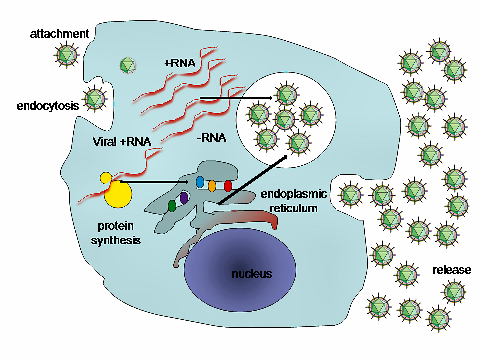 A typical virus replication cycle  Role in human disease Examples of common human diseases caused by viruses include the common cold, influenza, chickenpox and cold sores. Many serious diseases such as ebola, AIDS, avian influenza and SARS are caused by viruses. The relative ability of viruses to cause disease is described in terms of virulence. Other diseases are under investigation as to whether they too have a virus as the causative agent, such as the possible connection between human herpes virus six (HHV6) and neurological diseases such as multiple sclerosis and chronic fatigue syndrome. There is controversy over whether the borna virus, previously thought to cause neurological diseases in horses, could be responsible for psychiatric illnesses in humans.Viruses have different mechanisms by which they produce disease in an organism, which largely depends on the viral species. Mechanisms at the cellular level primarily include cell lysis, the breaking open and subsequent death of the cell. In multicellular organisms, if enough cells die, the whole organism will start to suffer the effects. Although viruses cause disruption of healthy homeostasis, resulting in disease, they may exist relatively harmlessly within an organism. An example would include the ability of the herpes simplex virus, which causes cold sores, to remain in a dormant state within the human body. This is called latency and is a characteristic of the herpes viruses including Epstein-Barr virus, which causes glandular fever, and varicella zoster virus, which causes chickenpox and shingles. Most people have been infected with at least one of these types of herpes virus. However, these latent viruses might sometimes be beneficial, as the presence of the virus can increase immunity against bacterial pathogens, such as Yersinia pestis.Some viruses can cause life-long or chronic infections, where the viruses continue to replicate in the body despite the host's defence mechanisms. This is common in hepatitis B virus and hepatitis C virus infections. People chronically infected are known as carriers, as they serve as reservoirs of infectious virus. In populations with a high proportion of carriers, the disease is said to be endemic
__________________
"Wa tu izzu man-ta shaa, wa tu zillu man-ta shaa"
|
|
«
Previous Thread
|
Next Thread
»
|
|

 Similar Threads
|
||||
| Thread | Thread Starter | Forum | Replies | Last Post |
| The miracle of the cell membrane | muhammadmohsinali | General Science & Ability | 0 | Tuesday, August 02, 2011 09:46 PM |
| Solved Everyday Science Papers | Dilrauf | General Science & Ability | 4 | Friday, April 08, 2011 06:10 PM |
| zoology paper 2 (part A) | dj don | Zoology | 0 | Friday, December 31, 2010 02:25 PM |
| Plant cell | Last Island | General Science Notes | 0 | Monday, August 06, 2007 09:40 PM |
| Animal cell | Last Island | General Science Notes | 0 | Sunday, August 05, 2007 05:50 AM |

