CSS Forums
Saturday, July 27, 2024
07:11 PM (GMT +5)
07:11 PM (GMT +5)


|
#71
 Monday, May 11, 2009
|
||||
|
||||
|
mashallah! u have a gr8 job......have u done masters in zoology?.....or its ur kind effort at ur own for css?
__________________
~Miss zoologist~ 
|


|
#72
Monday, May 11, 2009
|
|||
|
|||
|
Quote:
yes i have done masters in zoology  and M phil in zoology and M phil in zoology .this is for those who want to opt zoology .this is for those who want to opt zoology for css. for css.thanks and regards
|


| The Following User Says Thank You to AFRMS For This Useful Post: | ||
sunlight (Sunday, May 17, 2009) | ||
|
#73
Monday, May 11, 2009
|
|||
|
|||
|
Spicules Spicules are tiny spike-like structures of diverse origin and function found in many organisms, such as the copulatory spicules of certain nematodes or the grains on the skin of some frogs.This article discusses the skeletal spicules that occur in most sponges. They provide structural support and deter predators. Large spicules, visible to the naked eye are referred to as megascleres, while smaller, microscopic ones are termed microscleres. Spicules have four major symmetry types: Monaxon (simple cylinders with pointed ends), triaxon, tetraxon, and polyaxon. Sponges can be calcareous, siliceous, or composed of spongin. The meshing of many spicules serves as the sponges skeleton. The composition, size, and shape of spicules is one of the largest determining factors in sponge taxonomy. Spicules are formed by sclerocytes, which are derived from archaeocytes. The sclerocyte begins with an organic filament, and adds silica to it. Spicules are generally elongated at a rate of 1-10 μm per hour. Once the spicule reaches a certain length it protrudes from the sclerocyte cell body, but remains within the cells membrane. On occasion, sclerocytes may begin a second spicule while the first is still in progress. Research on the Euplectella aspergillum (Venus' Flower Basket) demonstrated that the spicules of certain deep-sea sponges have similar traits to Optical fibre. In addition to being able to trap and transport light, these spicules have a number of advantages over commercial fibre optic wire. They are stronger, resist stress easier, and form their own support elements. Also, the low-temperature formation of the spicules, as compared to the high temperature stretching process of commercial fibre optics, allows for the addition of impurities which improve the refractive index. In addition, these spicules have built-in lenses in the ends which gather and focus light in dark conditions. It has been theorized that this ability may function as a light source for symbioticalgae (as with Rosella racovitzae) or as an attractor for shrimp which live inside the Venus' Flower Basket. However, a conclusive decision has not been reached; it may be that the light capabilities are simply a coincidental trait from a purely structural element. 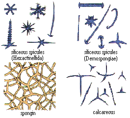 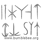
|
| The Following User Says Thank You to AFRMS For This Useful Post: | ||
sunlight (Sunday, May 17, 2009) | ||
|
#74
Tuesday, May 12, 2009
|
|||
|
|||
|
.................................................. .......................
|
| The Following User Says Thank You to AFRMS For This Useful Post: | ||
sunlight (Sunday, May 17, 2009) | ||
|
#75
Thursday, May 14, 2009
|
|||
|
|||
|
Germ layers
A germ layer is a group of cells, formed during animal embryogenesis. Germ layers are particularly pronounced in the vertebrates; however, all animals more complex than sponges (eumetazoans and agnotozoans) produce two or three primary tissue layers (sometimes called primary germ layers). Animals with radial symmetry, like cnidarians, produce two germ layers (the ectoderm and endoderm) making them diploblastic. Animals with bilateral symmetry produce a third layer between these two layers (appropriately called the mesoderm) making them triploblastic. Germ layers eventually give rise to all of an animals tissues and organs through the process of organogenesis. Among animals, sponges show the simplest organization, having a single germ layer. Although they have differentiated cells (e.g. collar cells), they lack true tissue coordination. Diploblastic animals, Cnidaria and ctenophores, show an increase in complexity, having two germ layers, the endoderm and ectoderm. Diploblastic animals are organized into recognisable tissues. All higher animals (from flatworms to humans) are triploblastic, possessing a mesoderm in additition to the germ layers found in Diploblasts. Triploblastic animals develop recognisable organs. Development Fertilization leads to the formation of a zygote. During the next stage, cleavage, mitotic cell divisions transform the zygote into a tiny ball of cells, a blastula. This early embryonic form undergoes gastrulation, forming a gastrula with either two or three layers (the germ layers). In all vertebrates, these are the forerunners of all adult tissues and organs. The appearance of the archenteron marks the onset of gastrulation. In humans, after about three days, the zygote forms a solid mass of cells by mitotic division, called a morula. This then changes to a blastocyst, consisting of an outer layer called a trophoblast, and an inner cell mass called the embryoblast. Filled with uterine fluid, the blastocyst breaks out of the zona pellucida and undergoes implantation. The inner cell mass initially has two layers: the hypoblast and epiblast. At the end of the second week, a primitive streak appears. The epiblast in this region moves towards the primitive streak, dives down into it, and forms a new layer, called the endoderm, pushing the hypoblast out of the way (this goes on to form the amnion.) The epiblast keeps moving and forms a second layer, the mesoderm. The top layer is now called the ectoderm. Endoderm The endoderm is one of the germ layers formed during animal embryogenesis. Cells migrating inward along the archenteron form the inner layer of the gastrula, which develops into the endoderm. The endoderm consists at first of flattened cells, which subsequently become columnar. It forms the epithelial lining of the whole of the digestive tube excepting part of the mouth and pharynx and the terminal part of the rectum (which are lined by involutions of the ectoderm). It also forms the lining cells of all the glands which open into the digestive tube, including those of the liver and pancreas; the epithelium of the auditory tube and tympanic cavity; the trachea, bronchi, and air cells of the lungs; the urinary bladder and part of the urethra; and the follicle lining of the thyroid gland and thymus. The endoderm forms: the stomach, the colon, the liver, the pancreas, the urinary bladder, the lining of the urethra, the epithelial parts of trachea, the lungs, the pharynx, the thyroid, the parathyroid, and the intestines. Mesoderm The mesoderm germ layer forms in the embryos of triploblasticanimals. During gastrulation, some of the cells migrating inward contribute to the mesoderm, an additional layer between the endoderm and the ectoderm. This key innovation evolved hundreds of millions of years ago and led to the evolution of nearly all large, complex animals. The formation of a mesoderm led to the development of a coelom. Organs formed inside a coelom can freely move, grow, and develop independently of the body wall while fluid cushions and protects them from shocks. The mesoderm forms: skeletal muscle, the skeleton, the dermis of skin, connective tissue, the urogenital system, the heart, blood (lymph cells), and the spleen. Ectoderm The ectoderm is the start of a tissue that covers the body surfaces. It emerges first and forms from the outermost of the germ layers. The ectoderm forms: the central nervous system, the lens of the eye, cranial and sensory, the ganglia and nerves, pigment cells, head connective tissues, the epidermis, hair, and mammary glands Neural crest Because of its great importance, the neural crest is sometimes considered a fourth germ layer. It is, however, derived from the ectoderm. 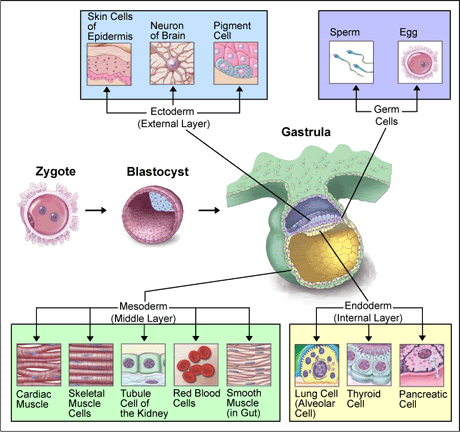 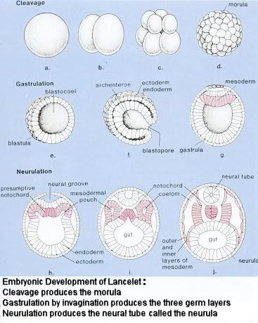 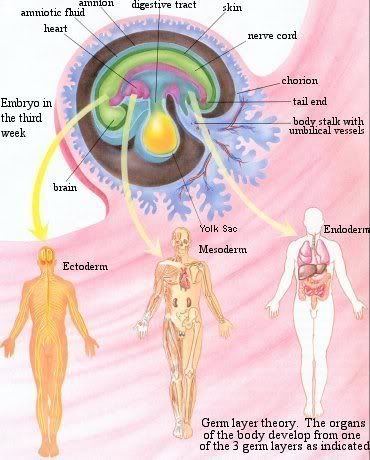 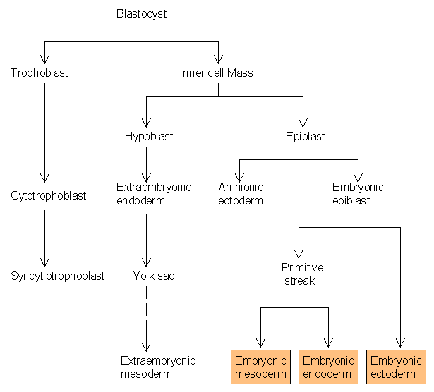
|
| The Following 2 Users Say Thank You to AFRMS For This Useful Post: | ||
prissygirl (Wednesday, September 02, 2009), sunlight (Sunday, May 17, 2009) | ||
|
#76
Thursday, May 14, 2009
|
|||
|
|||
|
Embryonic Membranes The embryos of reptiles, birds, and mammals produce 4 extraembryonic membranes, the amnion yolk sac chorion, and allantois In birds and most reptiles, the embryo with its extraembryonic membranes develops within a shelled egg. The amnion protects the embryo in a sac filled with amniotic fluid. The yolk sac contains yolk the sole source of food until hatching. Yolk is a mixture of proteins and lipoproteins. The chorion lines the inner surface of the shell (which is permeable to gases) and participates in the exchange of O2 and CO2 between the embryo and the outside air. The allantois stores metabolic wastes (chiefly uric acid) of the embryo and, as it grows larger, also participates in gas exchange. With these four membranes, the developing embryo is able to carry on essential metabolism while sealed within the egg. Surrounded by amniotic fluid, the embryo is kept as moist as a fish embryo in a pond. Although (most) mammals do not make a shelled egg, they do also enclose their embryo in an amnion. For this reason, the reptiles, birds, and mammals are collectively referred to as the amniota. Mammals fall into three groups that differ in the way they use the amniotic egg. Monotremes These primitive mammals produce a shelled egg like their reptilian ancestors. Only four species exist today: three species of spiny anteater (echidna) and the duckbill platypus Marsupial Marsupials do not produce a shelled egg. The egg, which is poorly supplied with yolk, is retained for a time within the reproductive tract of the mother. The embryo penetrates the wall of the uterus. The yolk sac provides a rudimentary connection to the mother's blood supply from which it receives food, oxygen, and other essentials. However, this interface between the tissues of the uterus and the extraembryonic membranes never becomes elaborately developed, and the young are born in a very immature state. Placental mammals In placental mammals, the extraembryonic membranes form a placenta and umbilical cord, which connect the embryo to the mother's uterus in a more elaborate and efficient way. The blood supply of the developing fetus is continuous with that of the placenta. The placenta extracts food and oxygen from the uterus. Carbon dioxide and other wastes (e.g., urea) are transferred to the mother for disposal by her excretory organs. Humans are placental mammals. 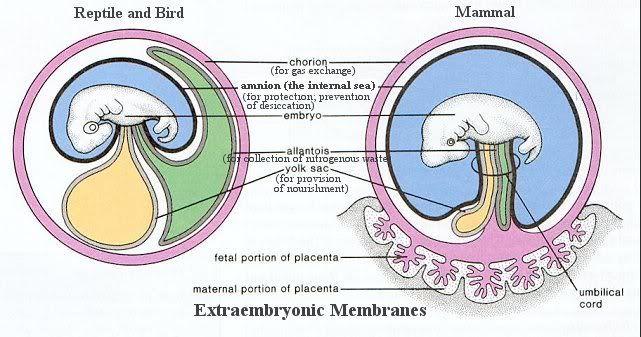 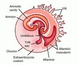 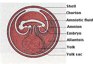
|
| The Following User Says Thank You to AFRMS For This Useful Post: | ||
sunlight (Sunday, May 17, 2009) | ||
|
#77
Friday, May 15, 2009
|
|||
|
|||
|
EGG TYPES The ZygoteThe zygote is totipotent - it has the potential to develop into any other cell Zygotes from different species differ in their yolk content: - oligolecithal eggs - little yolk - mesolecithal eggs - moderate amounts of yolk - macrolecithal eggs - large amounts of yolk The distribution of yolk also differs: - oligolecithal eggs tend to be isolecithal - yolk is distributed throughout the egg - meso- and macrolecithal eggs tend to be telolecithal - yolk is segregated toward the vegetal pole, away from the animal pole. 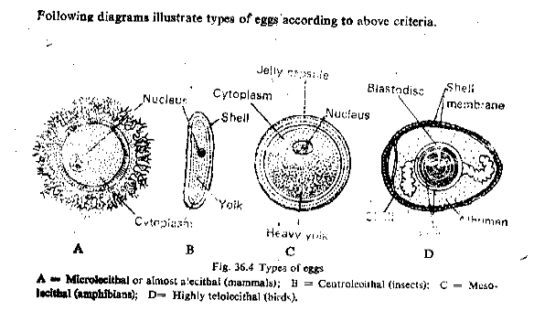 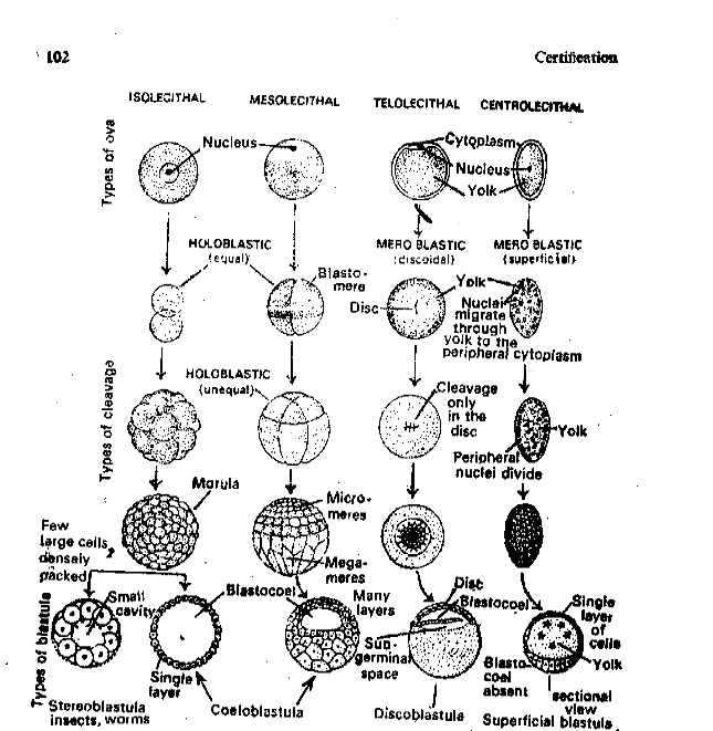 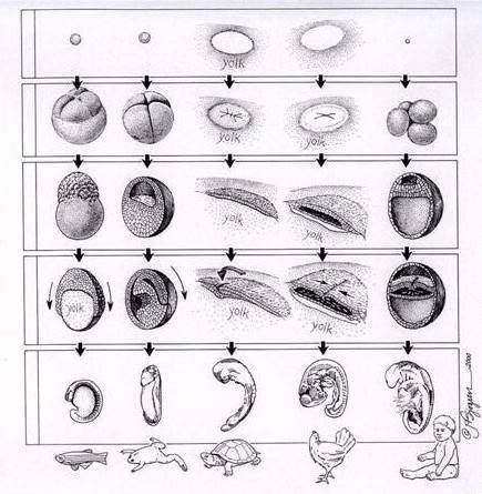 Cleavage Early cell division occurs without cell growth, and is termed cleavage The presence of yolk slows cleavage in proportion to its concentration - in holoblastic cleavage cell divisions are complete - in meroblastic cleavage, the yolk is not completely divided Cleavage results in the blastula - a mass of undifferentiated cells - cells have a high nucleus:cytoplasm ratio - cells are undifferentiated - the blastula contains a central cavity or blastocoel Gastrulation Gastrulation is the transformation of the blastula into a gastrula, a structure with three germ layers and a gastrocoel Gastrulation begins at the blastopore - a spot where proliferating cells fold into the blastocoel The gastrula has several important characters the three main body axes are defined cells acquire developmental fates - the ectoderm will form the epidermis, nervous system, and sense organs - the endoderm will form the gut lining and derivatives - the mesoderm will form everything else Neurulation Neurulation is the initial formation of the nervous system - growth, cell differentiation and organogenesis begin Chordamesoderm cells aggregate to form the notochord The chordamesoderm induces the dorsal ectoderm to form the neural tube - induction involves turning on or off of specific genetic pathways in one type of cell following contact by another type of cell Mesoderm flanking the notochord becomes segmented into laterally paired blocks or somites different regions of the somites and lateral mesoderm have different developmental fates Pharyngeal pouches (6-9) develop by evaginations of the pharyngeal endoderm, which meet indentations of the ectoderm - they give rise to several important structures: (e.g., the eardrum, jaws, glands) Placodes are ectodermal thickenings in the head - they contribute to the sense organs, and to the wandering cells Mesenchyme is the embryonic connective tissue - Wandering cells are mesenchyme cells that migrate through the embryo and form specific structures - Neural crest cells are wandering cells derived from ectoderm dorsal to the neural tube. - unique to vertebrates Organogenesis Organogenesis involves continued specialization of cells to form tissues - ectoderm and endoderm form predominantly epithelium - sheets with tight intercellular junctions, flanking open space - mesoderm forms the matrix of the organs, including a variety of tissues Tissues combine to form organs Organs unite into organ systems
|
| The Following User Says Thank You to AFRMS For This Useful Post: | ||
sunlight (Sunday, May 17, 2009) | ||
|
#78
Saturday, May 16, 2009
|
|||
|
|||
|
Differences between the hearts: Vertebrates The fish heart (figure 1a) is much different than the amphibian/reptile/bird/mammal heart (figures 1b and c). Hearts are very complex--they're not just a bunch of random arteries and veins connecting tissue. Fish hearts simply draw in deoxygenated blood in a single atrium, and pump it out through a ventricle. This system is termed "single circulation", as blood enters the heart, gets pumped through the gills and out to the body, Blood pressure is low for oxygenated blood leaving the gills. 3 and 4 chambered hearts have a pulmonary circuit (pathways taking blood from heart to lung and back to heart) that is very complex and must be set up such that blood can travel from the heart to become oxygenated in the lungs and then be properly pumped back the heart and out to the body. The 3 (and 4) chambered heart has "double circulation" (figure 1b and c) and is quite different from "single circulation" (figure 1a) of fishes. "Double circulation" has an interior circuit within the heart--blood enters the heart, leaves the heart and gets oxygenated, enters the heart again, and then gets pumped out to the body. Because "Double circulation" allows oxygenated blood to be pumped back into the heart before going out to the body, it pumps blood with much more pressure and much more vigorously than "single circulation". Though the 4 chambered heart has 2 atrium-ventricle pairs, both pairs do not do the same thing. There are 4 steps involved with blood entering the heart: 1) oxygen poor blood enters the first atrium. 2) oxygen poor blood is fed to the first ventricle, which pumps it out to the pulmonary circuit (lungs) where it is enriched in oxygen. 3) Oxygen rich blood just leaving the lungs is pumped back into the second atria. 4) Oxygen rich blood is then fed to the second ventricle, which pumps the oxygen rich blood out of the heart and back into the body for usage. The 4 chambered heart differs from the 3 chambered heart in that it keeps oxygenated blood completely separate from de-oxygnated blood, because there is one ventricle for deoxgynated blood and one for oxygenated blood. In the 3 chambered heart, a single ventricle pumps both out of the heart, and there is some mixing between fresh and old blood. The 2 ventricle-4 chamber heart prevents mixing allows the blood leaving the heart to have far more oxygen than it would otherwise. This is good for enhancing the more fast paced lifestyle that birds and mammals tend to have, giving an advantage to having a 4 chambered heart. 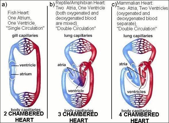
|
|
#79
Sunday, May 17, 2009
|
|||
|
|||
|
Hepatic portal system In human anatomy, the hepatic portal system is the system of veins comprised of the hepatic portal vein and its tributaries. It is also called the portal venous system, although it is not the only example of a portal venous system, and splanchnic veins, which is not synonymous with hepatic portal system and is imprecise (as it means visceral veins and not necessarily the veins of the abdominal viscera). Function The portal venous system is responsible for directing blood from parts of the gastrointestinal tract to the liver. Substances absorbed in the small intestine travel first to the liver for processing before continuing to the heart. Not all of the gastrointestinal tract is part of this system. The system extends from about the lower portion of the esophagus to the upper part of the anal canal. It also includes venous drainage from the spleen and pancreas. Many drugs that are absorbed through the GI tract are substantially metabolized by the liver before reaching general circulation. This is known as the first pass effect. As a consequence, certain drugs can only be taken via certain routes. For example, nitroglycerin cannot be swallowed because the liver would inactivate the medication, but it can be taken under the tongue or transdermal (through the skin) and thus is absorbed in a way that bypasses the portal venous system. Blood flow to the liver is unique in that it receives both oxygenated and deoxygenated blood. As a result, the partial pressure of oxygen (pO2) and perfusion pressure of portal blood are lower than in other organs of the body. Blood passes from branches of the portal vein through cavities between "plates" of hepatocytes called sinusoids. Blood also flows from branches of the hepatic artery and mixes in the sinusoids to supply the hepatocytes with oxygen. This mixture percolates through the sinusoids and collects in a central vein which drains into the hepatic vein. The hepatic vein subsequently drains into the inferior vena cava. Large veins that are considered part of the portal venous system are the: Hepatic portal vein Splenic vein Roughly, the portal venous system corresponds to areas supplied by the celiac trunk, the superior mesenteric artery, and the inferior mesenteric artery. 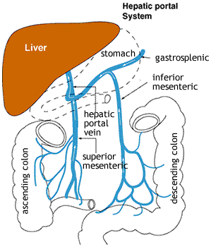 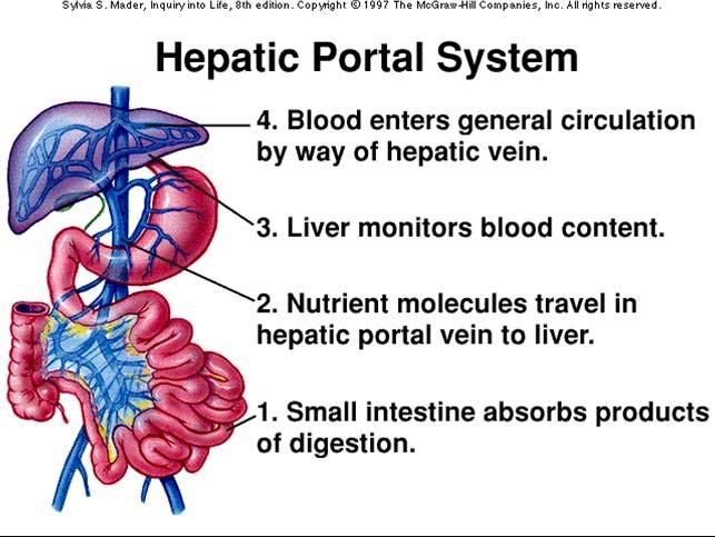 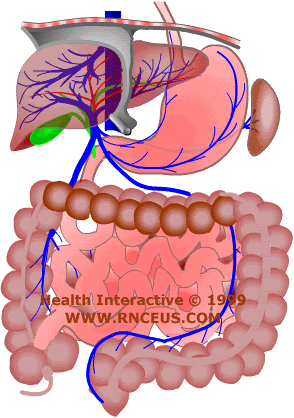 The hepatic portal system begins in the capillaries of the digestive organs and ends in the portal vein. Consequently, portal blood contains substances absorbed by the stomach and intestines. Portal blood is passed through the hepatic lobules where nutrients and toxins are absorbed, excreted or converted. Restriction of outflow through the hepatic portal system can lead to portal hypertension. Portal hypertension is most often associated with cirrhosis. Patients usually present with splenomegaly, ascites, GI bleeding and/or portal systemic encephalopathy. The consequences of portal hypertension are due to portal systemic anastomosis formed by the body as an attempt to bypass the obstructed liver circulation. These collateral vessels form along the falciform ligament, diaphragm, spleen, stomach and peritoneum. The collaterals find their way to the renal vein where blood drained from the digestive organs is let into the systemic circulation.
|
|
#80
Sunday, May 17, 2009
|
|||
|
|||
|
Blood Blood Components Normally, 7-8% of human body weight is from blood. In adults, this amounts to 4-5 quarts of blood. This essential fluid carries out the critical functions of transporting oxygen and nutrients to our cells and getting rid of carbon dioxide, ammonia, and other waste products. In addition, it plays a vital role in our immune system and in maintaining a relatively constant body temperature. Blood is a highly specialized tissue composed of many different kinds of components. Four of the most important ones are red cells, white cells, platelets, and plasma. All humans produce these blood components--there are no populational or regional differences. 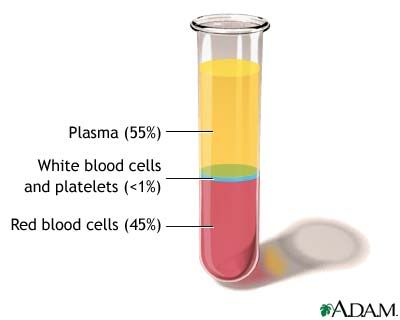 Red Cells Red cells, or erythrocytes , are relatively large microscopic cells without nuclei. In this latter trait, they are similar to the primitive prokaryotic cells of bacteria. Red cells normally make up 40-50% of the total blood volume. They transport oxygen from the lungs to all of the living tissues of the body and carry away carbon dioxide. The red cells are produced continuously in our bone marrow from stem cells at a rate of about 2-3 million cells per second. Hemoglobin is the gas transporting protein molecule that makes up 95% of a red cell. Each red cell has about 270,000,000 iron-rich hemoglobin molecules. People who are anemic generally have a deficiency in red cells. The red color of blood is primarily due to oxygenated red cells. Human fetal hemoglobin molecules differ from those produced by adults in the number of amino acid chains. Fetal hemoglobin has three chains, while adults produce only two. As a consequence, fetal hemoglobin molecules attract and transport relatively more oxygen to the cells of the body. 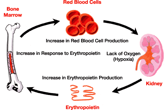 White Cells White cells, or leukocytes , exist in variable numbers and types but make up a very small part of blood's volume--normally only about 1% in healthy people. Leukocytes are not limited to blood. They occur elsewhere in the body as well, most notably in the spleen, liver, and lymph glands. Most are produced in our bone marrow from the same kind of stem cells that produce red blood cells. Others are produced in the thymus gland, which is at the base of the neck. Some white cells (called lymphocytes ) are the first responders for our immune system. They seek out, identify, and bind to alien protein on bacteria, viruses, and fungi so that they can be removed. Other white cells (called granulocytes and macrophages ) then arrive to surround and destroy the alien cells. They also have the function of getting rid of dead or dying blood cells as well as foreign matter such as dust and asbestos. Red cells remain viable for only about 4 months before they are removed from the blood and their components recycled in the spleen. Individual white cells usually only last 18-36 hours before they also are removed, though some types live as much as a year. The description of white cells presented here is a simplification. There are actually many specialized sub-types of them that participate in different ways in our immune responses. 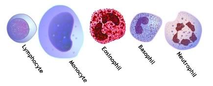 Platelets Platelets , or thrombocytes , are cell fragments without nuclei that work with blood clotting chemicals at the site of wounds. They do this by adhering to the walls of blood vessels, thereby plugging the rupture in the vascular wall. They also can release coagulating chemicals which cause clots to form in the blood that can plug up narrowed blood vessels. There are more than a dozen types of blood clotting factors and platelets that need to interact in the blood clotting process. Recent research has shown that platelets help fight infections by releasing proteins that kill invading bacteria and some other microorganisms. In addition, platelets stimulate the immune system. Individual platelets are about 1/3 the size of red cells. They have a lifespan of 9-10 days. Like the red and white blood cells, platelets are produced in bone marrow from stem cells. 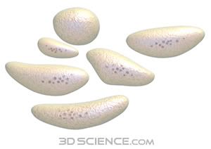 Plasma Plasma is the relatively clear liquid water (92+%), sugar, fat, protein and salt solution which carries the red cells, white cells, platelets, and some other chemicals. Normally, 55% of our blood's volume is made up of plasma. About 95% of it consists of water. As the heart pumps blood to cells throughout the body, plasma brings nourishment to them and removes the waste products of metabolism. Plasma also contains blood clotting factors, sugars, lipids, vitamins, minerals, hormones, enzymes, antibodies, and other proteins. It is likely that plasma contains some of every protein produced by the body--approximately 500 have been identified in human plasma so far.
|
 |
«
Previous Thread
|
Next Thread
»
|
|

 Similar Threads
|
||||
| Thread | Thread Starter | Forum | Replies | Last Post |
| Very Important : How to Prepare Study Notes | Shaa-Baaz | Tips and Experience Sharing | 5 | Sunday, May 21, 2017 08:30 PM |
| Effective Study Skills | Sureshlasi | Tips and Experience Sharing | 1 | Friday, November 16, 2007 09:28 AM |
| Regarding Notes | Anonymous84 | Tips and Experience Sharing | 1 | Wednesday, August 15, 2007 06:56 PM |

